. 2021 Apr 1;14(4):307.
doi: 10.3390/ph14040307.

Novel Mechanism for Memantine in Attenuating Diabetic Neuropathic Pain in Mice via Downregulating the Spinal HMGB1/TRL4/NF-kB Inflammatory Axis

Affiliations

Novel Mechanism for Memantine in Attenuating Diabetic Neuropathic Pain in Mice via Downregulating the Spinal HMGB1/TRL4/NF-kB Inflammatory Axis

Suliman Y Alomar et al. Pharmaceuticals (Basel). .

Abstract

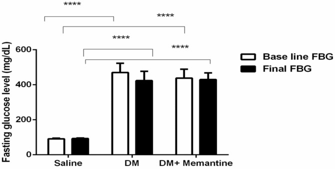
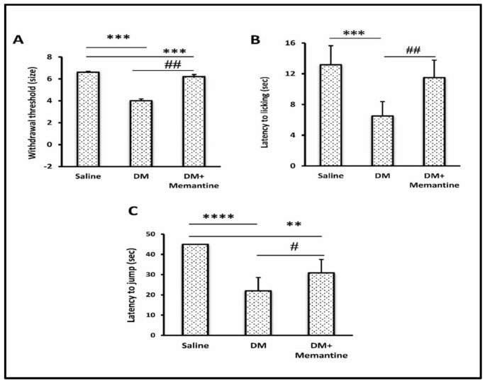
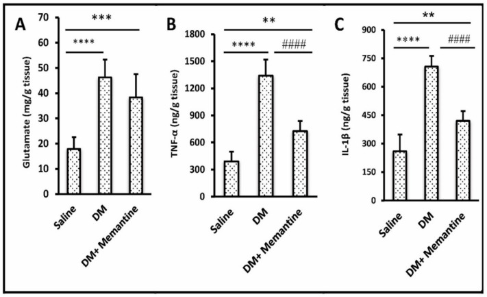
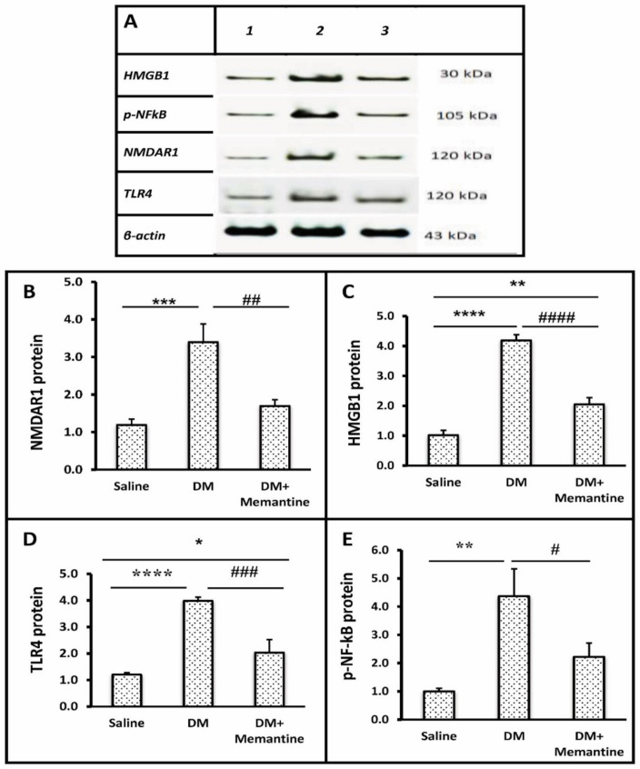
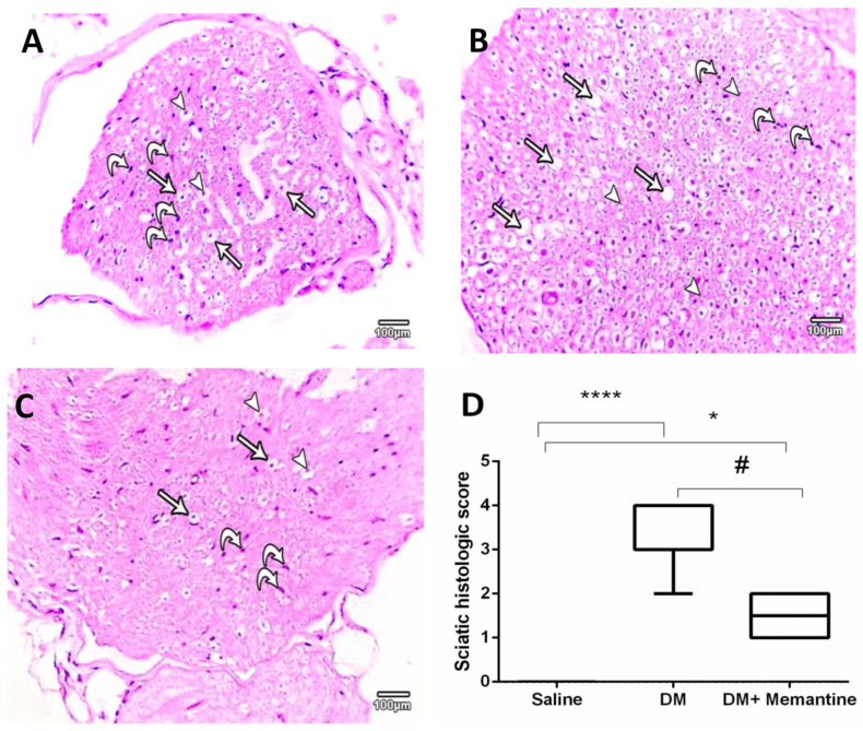
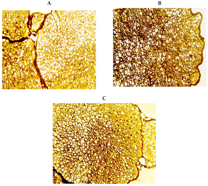
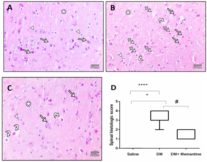
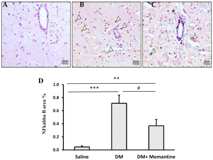
Diabetic neuropathic pain (DNP) is a common diabetic complication that currently lacks an efficient therapy. The aim of the current work was to uncover the anti-allodynic and neuroprotective effects of memantine in a model of mouse diabetic neuropathy and its ameliorative effect on the high-mobility group box-1 (HMGB1)/toll-like receptor 4 (TLR4)/nuclear factor-k B (NF-kB) inflammatory axis. Diabetes was prompted by an alloxan injection (180 mg/kg) to albino mice. On the ninth week after diabetes induction, DNP was confirmed. Diabetic mice were randomly allocated to two groups (six mice each); a diabetes mellitus (DM) group and DM+memantine group (10 mg/kg, daily) for five weeks. DNP-related behaviors were assessed in terms of thermal hyperalgesia and mechanical allodynia by hot-plate and von Frey filaments. Enzyme-linked immunosorbent assay (ELISA) kits were used to measure the spinal glutamate, interleukin-1 beta (IL-1β), and tumor necrosis factor-α (TNF-α). The spinal levels of N-methyl-D-aspartate type 1 receptor (NMDAR1), HMGB1, TLR4, and phosphorylated NF-kB were assessed using Western blotting. Histopathological investigation of the spinal cord and sciatic nerves, together with the spinal cord ultrastructure, was employed for assessment of the neuroprotective effect. Memantine alleviated pain indicators in diabetic mice and suppressed excessive NMDAR1 activation, glutamate, and pro-inflammatory cytokine release in the spinal cord. The current study validated the ability of memantine to combat the HMGB1/TLR4/NF-kB axis and modulate overactive glutamate spinal transmission, corroborating memantine as an appealing therapeutic target in DNP.

Keywords: HMGB1/TRL4/NF-kB axis; glutamate; memantine; mouse diabetic neuropathy; sciatic pathology.


Conflict of interest statement

The authors declare no conflict of interest.

Figures

Figure 1
Figure 1
Effect of memantine on the blood glucose level. Fasting blood glucose was measured before memantine therapy and at the end of the experiment. The data are the mean ± standard deviation of the mean (SDM) and were analyzed by one-way ANOVA and Tukey’s tests. DM: diabetes mellitus mice, **** p < 0.0001 versus the saline group.
Figure 2
Figure 2
Effect of memantine on allodynia and hyperalgesia. Diabetic mice were tested for (A) the von Frey test, (B) latency to licking (sec) and (C) latency to jump. Data are the mean ± SDM and analyzed by one-way ANOVA and Tukey’s tests. DM: diabetes mellitus mice, SDM: standard deviation of the mean. ** p < 0.01, *** p < 0.001, and **** p < 0.0001, versus the saline group; # p < 0.05 and ## p < 0.01 versus the DM group.
Figure 3
Figure 3
Effect of memantine on spinal glutamate and inflammatory mediators. Spinal cord samples were analyzed for (A) glutamate, (B) TNF-α, and (C) IL-1β. Data are the mean ± SDM and analyzed by one-way ANOVA and Tukey’s tests. DM: diabetes mellitus mice, and SDM: standard deviation of the mean, TNF-α: tumor necrosis factor-α, IL-1β: interlukin-1β. ** p < 0.01, *** p < 0.001, and **** p < 0.0001, versus the saline group; #### p < 0.0001 versus the DM group.
Figure 4
Figure 4
Effect of memantine on the spinal protein level of the inflammatory markers. (A) Spinal cord samples from saline (#1), diabetic (#2), and DM+memantine (10 mg/kg) groups (#3) were analyzed for (B) NMDAR1, (C) HMGB1, (D) TLR4 and (E) p-NF-kB. Data are mean ± SDM and analyzed by one-way ANOVA and Tukey’s tests. DM: diabetes mellitus mice, NMDAR1: n-methyl-D-aspartate receptors, HMGB1: high-mobility group protein 1, p-NF-kB: phosphorylated nuclear factor-kappa B, TLR4: Toll-like receptor 4, and SDM: standard deviation of the mean. * p < 0.05, ** p < 0.01, *** p < 0.001, and **** p < 0.0001 versus the saline group; # p < 0.05, ## p < 0.01, ### p < 0.001, and #### p < 0.0001 versus the DM group.
Figure 5
Figure 5
Microscopic pictures of hematoxylin and eosin stained sections of sciatic nerves. (A) An image from the saline group presenting multiple axons (arrowheads) found in different thicknesses of myelin sheathes (arrows) and enclosed within the endoneurium with multiple nuclei of Schwann cells (curved arrows). (B) Diabetic group (DM) showing several lost axons (arrows) with lost myelin sheathes (arrowheads) with some Schwann cell nuclei (curved arrows). (C DM+memantine shows normal neuronal axons (arrowheads) with normal myelin sheath (arrows) and an increased number of Schwann cells nuclei (curved arrows). ×100 bar 100 μm. (D)) Data from histopathologic scoring of the sciatic nerve sections as a box-whisker plot demonstrating the median values and analyzed by Kruskal–Wallis ANOVA. * p < 0.05, and **** p < 0.0001, versus the saline group; # p < 0.05 versus the DM group.
Figure 6
Figure 6
Silver staining for transverse sciatic nerve specimens. Images show well organized nerve fiber axons, including well-structured myelin sheathes enclosed by endoneurium in the saline group (A), profound staining by silver stain and a substantial decrease in the myelin sheathes in the diabetic group (B), less degeneration in the myelin sheathes and reduced silver staining in the DM+memantine group (C). Silver stain ×400.
Figure 7
Figure 7
Microscopic pictures of hematoxylin and eosin stained cross sections of spinal cord. (A) Saline group showing normal neural cells (arrows) and glia cells (arrow heads) surrounded by neuropils (star). (B) Diabetic group (DM) showing marked neural cell degeneration (arrows), vacuoles in the neuropils (crossed arrow), dilated capillaries (curved arrows), and gliosis (arrow head). (C) DM+memantine group showing preserved neural cells (arrows) and glial cells (arrow head) with reduced vacuolation in the neuropil (curved arrows). ×100; bar 100 µm. (D) box-plot demonstrating the medians of the histopathologic scores assigned for the spinal cord specimens. Analysis was done using Kruskal–Wallis ANOVA. * p < 0.05, and **** p < 0.0001 versus the saline group; # p < 0.05 versus the DM group.
Figure 8
Figure 8
Microscopic pictures of NF-kB immunostained sections from the spinal cords. Images show cross sections with: (A) very mild staining in the saline group, (B) strong positive nuclear expression in neural cells (arrow heads) from the diabetic group, and (C) decreased positive nuclear expression in neural cells (arrow heads) from the DM+memantine group. Immunohistochemistry counterstained with Mayer’s hematoxylin. ×100; bar 100 μm. (D) Data are mean staining area ± SDM. DM: diabetes mellitus, SDM: standard deviation of the mean, and NF-kB: nuclear factor-kappa B. ** p < 0.01, and *** p < 0.001 versus the saline group; # p < 0.05 versus the DM group.
Figure 9
Figure 9
Transmission electron microscopy images of sections of the spinal cord. Images in groups; control, diabetic, and treated diabetic group. Panel (A): control section showing the normal characteristics of myelinated axons with regular myelin sheathes (arrows). Panel (B1,B2) diabetic sections showing degenerated myelinated axons (head arrows) and phagocytic microglia/macrophage cells (M) with degenerating myelin and phagocytosed matter in the cytoplasm in the white matter. The degenerated axons showing disorganized myelin sheathes with swollen vacuolated axoplasm. Microglia are surrounding the degenerated axon of a collapsed myelin sheath. Panel (C) Treated group showing myelinated axons with regular myelin sheathes, and very small unmyelinated axons were found with thickened Schwan cell basement membranes (arrows).

References

    1. Ochoa J.L. Neuropathic pain: Redefinition and a grading system for clinical and research purposes. Neurology. 2009;72:1282–1283. doi: 10.1212/01.wnl.0000346325.50431.5f. - DOI - PubMed
    1. Bril V., England J., Franklin G.M., Backonja M., Cohen J., Del Toro D., Feldman E., Iverson D.J., Perkins B., Russell J.W., et al. Evidence-based Guideline: Treatment of Painful Diabetic Neuropathy: Report of the American Academy of Neurology, the American Association of Neuromuscular and Electrodiagnostic Medicine, and the American Academy of Physical Medicine and Rehabilitation. PMR. 2011;3:345–352. doi: 10.1016/j.pmrj.2011.03.008. - DOI - PubMed
    1. Brod M., Pohlman B., Blum S.I., Ramasamy A., Carson R. Burden of Illness of Diabetic Peripheral Neuropathic Pain: A Qualitative Study. Patient Patient-Cent. Outcomes Res. 2014;8:339–348. doi: 10.1007/s40271-014-0093-9. - DOI - PubMed
    1. Obrosova I.G. Diabetic painful and insensate neuropathy: Pathogenesis and potential treatments. Neurotherapeutics. 2009;6:638–647. doi: 10.1016/j.nurt.2009.07.004. - DOI - PMC - PubMed
    1. Rosenberger D.C., Blechschmidt V., Timmerman H., Wolff A., Treede R.-D. Challenges of neuropathic pain: Focus on diabetic neuropathy. J. Neural Transm. 2020;127:589–624. doi: 10.1007/s00702-020-02145-7. - DOI - PMC - PubMed

LinkOut - more resources