

Multipotent Stromal Cell Extracellular Vesicle Distribution in Distant Organs after Introduction into a Bone Tissue Defect of a Limb
- PMID: 33916128
- PMCID: PMC8066794
- DOI: 10.3390/life11040306
Multipotent Stromal Cell Extracellular Vesicle Distribution in Distant Organs after Introduction into a Bone Tissue Defect of a Limb
Abstract
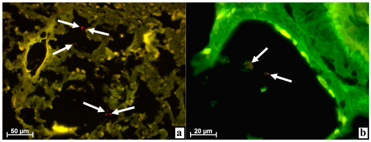
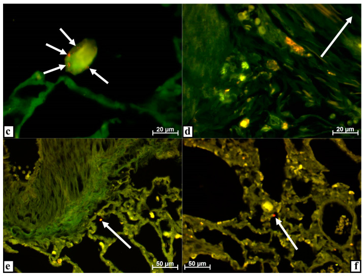
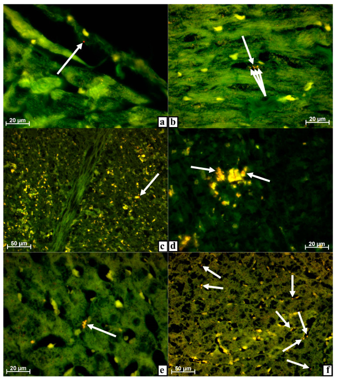
When administered intravenously, extracellular vesicles derived from multipotent stromal cells (MSC EVs) immediately pass through the lungs along with the blood and regularly spread to all organs. When administered intraperitoneally, they are absorbed either into the blood or into the lymph and are quickly disseminated throughout the body. The possibility of generalized spread of MSC EVs to distant organs in case of local intratissular administration remains unexplored. However, it is impossible to exclude MSC EV influence on tissues distant from the injection site due to the active or passive migration of these injected nanoparticles through the vessels. The research is based on findings obtained when studying the samples of lungs, heart, spleen, and liver of outbred rabbits of both sexes weighing 3-4 kg at various times after the injection of EVs derived from MSCs of bone marrow origin and labeled by PKH26 into an artificially created defect of the proximal condyle of the tibia. MSC EVs were isolated by serial ultracentrifugation and characterized by transmission electron microscopy and flow cytometry. After the introduction of MSC EVs into the damaged proximal condyle of the tibia of rabbits, these MSC EVs can be found most frequently in the lungs, myocardium, liver, and spleen. MSC EVs enter all of these organs with the blood flow. The lungs contained the maximum number of labeled MSC EVs; moreover, they were often associated with detritus and were located in the lumen of the alveoli. In the capillary network of various organs except the myocardium, MSC EVs are adsorbed by paravasal phagocytes; in some cases, specifically labeled small dust-like objects can be detected throughout the entire experiment-up to ten days of observation. Therefore, we can conclude that the entire body, including distant organs, is effected both by antigenic detritus, which appeared in the bloodstream after extensive surgery, and MSC EVs introduced from the outside.
Keywords: bone tissue defect of a limb; exosomes; extracellular vesicles distribution; heart; liver; lungs; multipotent stromal cell extracellular vesicles; spleen.
Conflict of interest statement
The authors declare that they have no conflict of interest. The sponsors had no role in the design, execution, interpretation, or writing of the study.
Figures
References
-
- Assis A.C., Carvalho J.L., Jacoby B.A., Ferreira R.L., Castanheira P., Diniz S.O., Cardoso V.N., Goes A.M., Ferreira A.J. Time-dependent migration of systemically delivered bone marrow mesenchymal stem cells to the infarcted heart. Cell Transplant. 2010;19:219–230. doi: 10.3727/096368909X479677. - DOI - PubMed
-
- Maiborodin I.V., Maslov R.V., Mikheeva T.V., Khomenyuk S.V., Maiborodina V.I., Morozov V.V., Ryaguzov M.E., Marchukov S.V., Kozlova J.N. The distribution of multipotent mesenchymal stromal cells and their detritus throughout the organism after subcutaneous introduction. Zhurnal Obshchei Biologii. 2020;81:96–107. doi: 10.31857/S0044459620020050. - DOI
-
- Maiborodin I.V., Maslov R.V., Mikheeva T.V., Marchukov S.V., Maiborodina V.I., Shevela A.A. Opportunity for elimination of injected multipotent stromal cells via lungs. Cell. Ther. Transpl. 2020;9:67–73. doi: 10.18620/ctt-1866-8836-2020-9-1-67-73. - DOI
-
- Maiborodin I.V., Maslov R.V., Mikheeva T.V., Elovskiy A.A., Figurenko N.F., Maiborodina V.I., Shevela A.I., Anishchenko V.V. The possibility of the angiogenesis in tissues remote from the place of the multipotent mesenchymal stromal cell injection. Molekulyarnaya Meditsina. 2018;16:22–26. doi: 10.29296/24999490-2018-03-04. - DOI
LinkOut - more resources
Full Text Sources
Other Literature Sources
