

Submandibular Push Exercise Using Visual Feedback from a Pressure Sensor in Patients with Swallowing Difficulties: A Pilot Study
- PMID: 33916285
- PMCID: PMC8065833
- DOI: 10.3390/healthcare9040407
Submandibular Push Exercise Using Visual Feedback from a Pressure Sensor in Patients with Swallowing Difficulties: A Pilot Study
Abstract
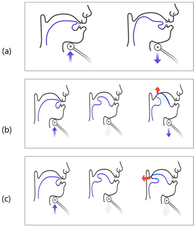
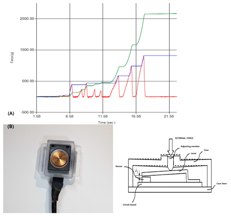
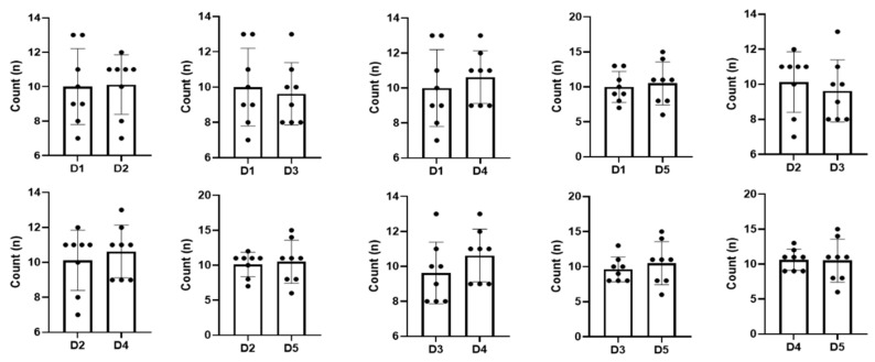
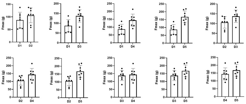
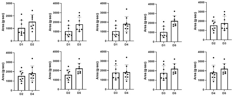
Objectives: We aimed to determine the usefulness and effectiveness of a submandibular push exercise with visual feedback from a pressure sensor in patients with dysphagia through continuous exercise sessions. Methods: Twelve patients with dysphagia of various etiologies were included. A total of five exercise sessions (every 3 or 4 days) over three weeks were conducted. During the submandibular push exercise, patients were instructed to maintain a maximum force for 3 s, repeated for 1 min to measure the number of exercises, the maximum pressure, and the area of the pressure-time graph. We statistically compared the values of each exercise trial. Results: Among the 12 patients, eight completed the exercise sessions. As the number of exercise trials increased, the maximum pressure and the area in the pressure-time graph showed a significant increase compared to the previous attempt (p < 0.05). The maximum pressure and the area of the pressure-time graph improved from the first to the fourth session (p < 0.05). The values were maintained after the fourth session, and there was no significant difference between the fourth and the fifth exercise (p > 0.05). There was no significant difference between successful and non-successful groups, except for the Modified Barthel Index (p < 0.05). Conclusion: Through repetitive exercise training, the submandibular push exercise using visual feedback from a pressure sensor can be applied as an exercise method to strengthen swallowing related muscles, such as the suprahyoid and infrahyoid muscles. However, additional studies including more patients and a long-term study period are warranted to evaluate the effects of the exercise for improvement of dysphagia.
Keywords: CTAR; Shaker exercise; infrahyoid; submandibular push exercise; suprahyoid.
Conflict of interest statement
The authors declare no conflict of interest.
Figures
References
-
- Park D., Lee H.H., Lee S.T., Oh Y., Lee J.C., Nam K.W., Ryu J.S. Normal contractile algorithm of swallowing related muscles revealed by needle EMG and its comparison to videofluoroscopic swallowing study and high resolution manometry studies: A preliminary study. J. Electromyogr. Kinesiol. 2017;36:81–89. doi: 10.1016/j.jelekin.2017.07.007. - DOI - PubMed
LinkOut - more resources
Full Text Sources
Other Literature Sources
Research Materials
