

Kidney Injury Caused by Preeclamptic Pregnancy Recovers Postpartum in a Transgenic Rat Model
- PMID: 33916404
- PMCID: PMC8038582
- DOI: 10.3390/ijms22073762
Kidney Injury Caused by Preeclamptic Pregnancy Recovers Postpartum in a Transgenic Rat Model
Abstract
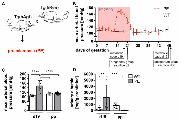
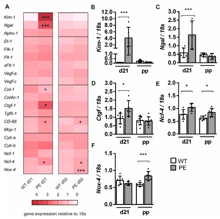
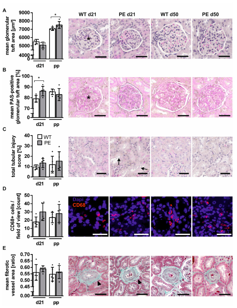
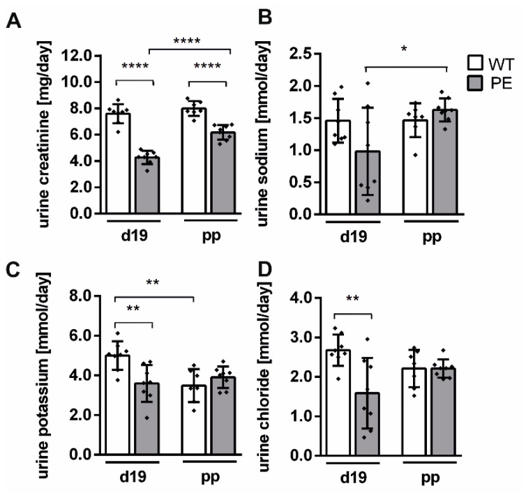
Preeclampsia (PE) is characterized by the onset of hypertension (≥140/90 mmHg) and presence of proteinuria (>300 mg/L/24 h urine) or other maternal organ dysfunctions. During human PE, renal injuries have been observed. Some studies suggest that women with PE diagnosis have an increased risk to develop renal diseases later in life. However, in human studies PE as a single cause of this development cannot be investigated. Here, we aimed to investigate the effect of PE on postpartum renal damage in an established transgenic PE rat model. Female rats harboring the human-angiotensinogen gene develop a preeclamptic phenotype after mating with male rats harboring the human-renin gene, but are normotensive before and after pregnancy. During pregnancy PE rats developed mild tubular and glomerular changes assessed by histologic analysis, increased gene expression of renal damage markers such as kidney injury marker 1 and connective-tissue growth factor, and albuminuria compared to female wild-type rats (WT). However, four weeks postpartum, most PE-related renal pathologies were absent, including albuminuria and elevated biomarker expression. Only mild enlargement of the glomerular tuft could be detected. Overall, the glomerular and tubular function were affected during pregnancy in the transgenic PE rat. However, almost all these pathologies observed during PE recovered postpartum.
Keywords: kidney injury; postpartum; preeclampsia; transgenic rat model.
Conflict of interest statement
The authors declare no conflict of interest.
Figures
Similar articles
-
Renin-angiotensin system transgenic mouse model recapitulates pathophysiology similar to human preeclampsia with renal injury that may be mediated through VEGF.Am J Physiol Renal Physiol. 2017 Mar 1;312(3):F445-F455. doi: 10.1152/ajprenal.00108.2016. Epub 2016 Dec 7. Am J Physiol Renal Physiol. 2017. PMID: 27927648
-
Renal glomerular and tubular injury in the offspring of the preeclampsia-like syndrome.Sci Rep. 2025 Jan 6;15(1):915. doi: 10.1038/s41598-025-85258-x. Sci Rep. 2025. PMID: 39762506 Free PMC article.
-
Systemic Outcomes of (Pyr1)-Apelin-13 Infusion at Mid-Late Pregnancy in a Rat Model with Preeclamptic Features.Sci Rep. 2019 Jun 12;9(1):8579. doi: 10.1038/s41598-019-44971-0. Sci Rep. 2019. PMID: 31189936 Free PMC article.
-
Glomerular disturbances in preeclampsia: disruption between glomerular endothelium and podocyte symbiosis.Hypertens Pregnancy. 2010 Jan;29(1):10-20. doi: 10.3109/10641950802631036. Hypertens Pregnancy. 2010. PMID: 19263286 Review.
-
The glomerular injury of preeclampsia.J Am Soc Nephrol. 2007 Aug;18(8):2281-4. doi: 10.1681/ASN.2007020255. Epub 2007 Jul 18. J Am Soc Nephrol. 2007. PMID: 17634433 Review.
Cited by
-
Computational model captures cardiac growth in hypertensive pregnancies and in the postpartum period.Am J Physiol Heart Circ Physiol. 2024 Jun 1;326(6):H1491-H1497. doi: 10.1152/ajpheart.00104.2024. Epub 2024 Apr 26. Am J Physiol Heart Circ Physiol. 2024. PMID: 38668702 Free PMC article.
-
Guidelines for assessing maternal cardiovascular physiology during pregnancy and postpartum.Am J Physiol Heart Circ Physiol. 2024 Jul 1;327(1):H191-H220. doi: 10.1152/ajpheart.00055.2024. Epub 2024 May 17. Am J Physiol Heart Circ Physiol. 2024. PMID: 38758127 Free PMC article. Review.
-
Pathogenesis of Pregnancy-Related Complications 1.0 and 2.0.Int J Mol Sci. 2022 Mar 11;23(6):3020. doi: 10.3390/ijms23063020. Int J Mol Sci. 2022. PMID: 35328441 Free PMC article.
-
Renal functional, transcriptome, and methylome adaptations in pregnant Sprague Dawley and Brown Norway rats.PLoS One. 2022 Jun 16;17(6):e0269792. doi: 10.1371/journal.pone.0269792. eCollection 2022. PLoS One. 2022. PMID: 35709218 Free PMC article.
References
-
- Brown M.A., Magee L.A., Kenny L.C., Karumanchi S.A., McCarthy F.P., Saito S., Hall D.R., Warren C.E., Adoyi G., Ishaku S. Hypertensive Disorders of Pregnancy: ISSHP Classification, Diagnosis, and Management Recommendations for International Practice. Hypertension. 2018;72:24–43. doi: 10.1161/HYPERTENSIONAHA.117.10803. - DOI - PubMed
-
- ACOG Practice Bulletin No. 202. American College of Obstetricians and Gynecologists. Gestational hypertension and preeclampsia. Obstet. Gynecol. 2019;133:211–214. doi: 10.1097/AOG.0000000000003019. - DOI
MeSH terms
Grants and funding
LinkOut - more resources
Full Text Sources
Other Literature Sources
Medical
