. 2021 Apr 5;10(4):810.
doi: 10.3390/cells10040810.

Inhibition of Cochlear HMGB1 Expression Attenuates Oxidative Stress and Inflammation in an Experimental Murine Model of Noise-Induced Hearing Loss

Affiliations

Inhibition of Cochlear HMGB1 Expression Attenuates Oxidative Stress and Inflammation in an Experimental Murine Model of Noise-Induced Hearing Loss

Cheng-Ping Shih et al. Cells. .

Abstract

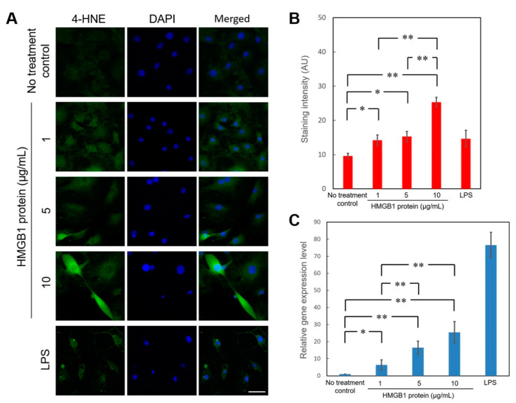
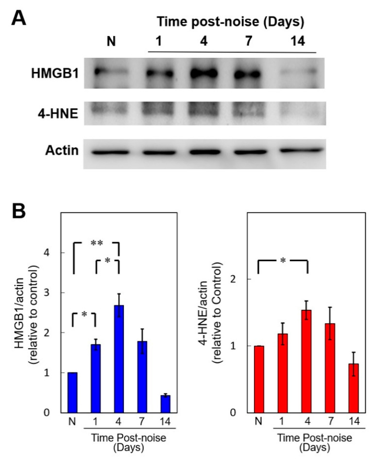
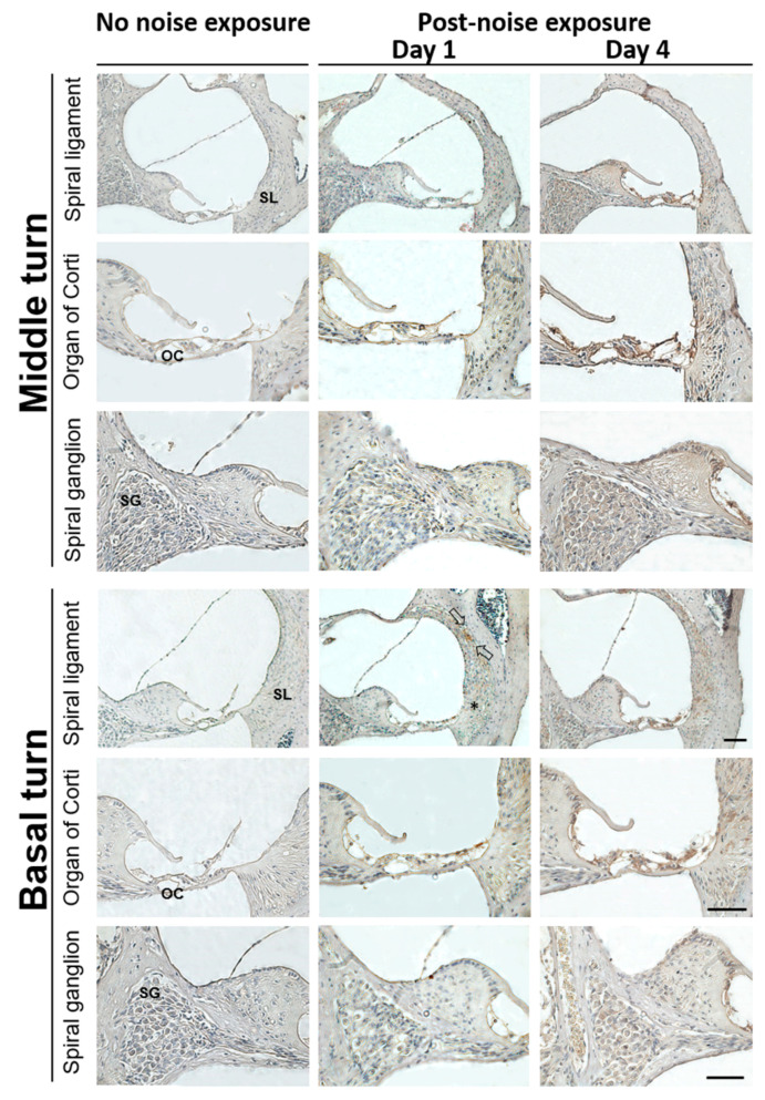
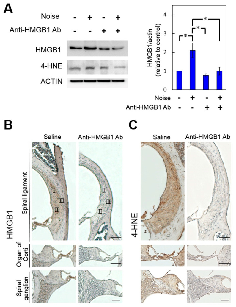
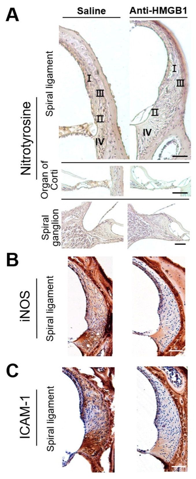
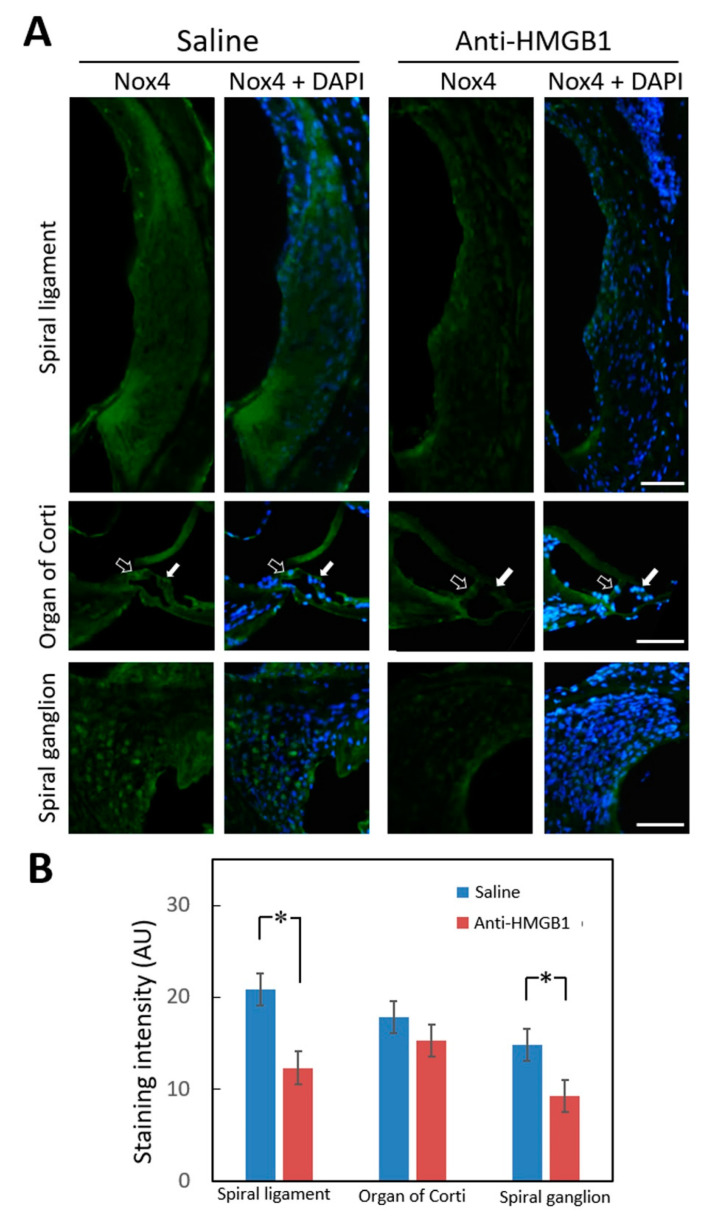
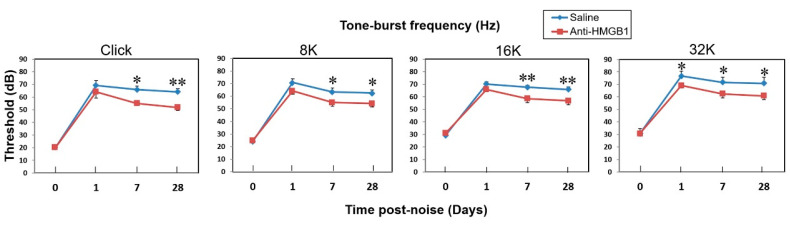
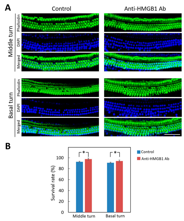
Noise-induced hearing loss (NIHL) is a common inner ear disease but has complex pathological mechanisms, one of which is increased oxidative stress in the cochlea. The high-mobility group box 1 (HMGB1) protein acts as an inflammatory mediator and shows different activities with redox modifications linked to the generation of reactive oxygen species (ROS). We aimed to investigate whether manipulation of cochlear HMGB1 during noise exposure could prevent noise-induced oxidative stress and hearing loss. Sixty CBA/CaJ mice were divided into two groups. An intraperitoneal injection of anti-HMGB1 antibodies was administered to the experimental group; the control group was injected with saline. Thirty minutes later, all mice were subjected to white noise exposure. Subsequent cochlear damage, including auditory threshold shifts, hair cell loss, expression of cochlear HMGB1, and free radical activity, was then evaluated. The levels of HMGB1 and 4-hydroxynonenal (4-HNE), as respective markers of reactive nitrogen species (RNS) and ROS formation, showed slight increases on post-exposure day 1 and achieved their highest levels on post-exposure day 4. After noise exposure, the antibody-treated mice showed markedly less ROS formation and lower expression of NADPH oxidase 4 (NOX4), nitrotyrosine, inducible nitric oxide synthase (iNOS), and intercellular adhesion molecule-1 (ICAM-1) than the saline-treated control mice. A significant amelioration was also observed in the threshold shifts of the auditory brainstem response and the loss of outer hair cells in the antibody-treated versus the saline-treated mice. Our results suggest that inhibition of HMGB1 by neutralization with anti-HMGB1 antibodies prior to noise exposure effectively attenuated oxidative stress and subsequent inflammation. This procedure could therefore have potential as a therapy for NIHL.

Keywords: NADPH oxidase (NOX); cochlea; high-mobility group box 1 (HMGB1); inflammation; noise-induced hearing loss (NIHL); oxidative stress; reactive nitrogen species (RNS); reactive oxygen species (ROS).


Conflict of interest statement

The authors declare no conflict of interest.

Figures

Figure 1
Figure 1
Recombinant HMGB1 activated 4-HNE production and induced the expression of iNOS gene in primary cochlear cells. (A) After incubation with various concentrations of recombinant HMGB1 for 24 h, immunostaining for 4-HNE was used to determine the generation of reactive oxygen species in primary cochlear cells. Representative immunofluorescence staining for 4-HNE (green), DAPI (blue), and merged images in the cells treated with recombinant HMGB1 or LPS. (B) Histogram representations of mean fluorescence intensity of 4-HNE staining intensities. Data are shown as the means ± SEM (n = 6 for each bar). Scale bars = 50 μm. (C) Recombinant HMGB1 stimulated the expression of iNOS gene (NOS2) in primary cochlear cells. Gene expression level was determined by quantitative PCR and expressed as the level relative to no treatment controls. Data are shown as the means ± SEM (n = 5 for each bar). * p < 0.05; ** p < 0.01; 4-HNE = 4-Hydroxynonenal; DAPI = 4,6-diamidino-2-phenylindole; LPS = lipopolysaccharide; SEM = standard error of the mean.
Figure 2
Figure 2
Noise exposure upregulates cochlear expression of high mobility group box 1 (HMGB1) and 4-HNE. (A) Western blot analysis of cochlear HMGB1 and 4-HNE expression after noise exposure. (B) Quantification of the time course of cochlear HMGB1 and 4-HNE expression (n = 4 [refers to 8 cochleae from 4 animals] for each bar). The results are expressed as the mean ± SEM. * p < 0.05; ** p < 0.01; N = a control mouse group not exposed to noise; SEM = standard error of the mean.
Figure 3
Figure 3
Representative expression and distribution of HMGB1 in mouse cochlear tissues following noise exposure. HMGB1 immunohistochemical staining (brown color) was substantially increased on post-exposure day 1 in the spiral ligament of the basal turn. Arrows indicate HMGB1-positive staining cells, mainly localized in the type II cell region. On day 4, the HMGB1 was markedly expressed in the organ of Corti, spiral limbus, spiral ganglion, and spiral ligament of the cochlear basal and middle turns (n = 4 [refers to 4 cochleae from 4 different animals]). Scale bars = 50 μm. SL = spiral ligament; OC = organ of Corti; SG = spiral ganglion.
Figure 4
Figure 4
Blockade of HMGB1 by pre-treatment with anti-HMGB1 antibodies diminished the noise-induced increases in ROS in the cochlea. Mice were intraperitoneally treated with anti-HMGB1 antibodies or saline 30 min prior to noise exposure. Samples of cochlear homogenates or sections were collected 4 days after noise exposure. (A) Western blot analysis for HMGB1 and 4-HNE in the cochleae treated with anti-HMGB1 antibodies or saline (n = 4 [refers to 8 cochleae from 4 different animals] for each bar). Representative immunohistochemical staining (brown color) for (B) HMGB1 and (C) 4-HNE of the cochleae (n = 4 [refers to 4 cochleae from 4 different animals]). Scale bars = 50 μm. * p < 0.05.
Figure 5
Figure 5
Blockade of HMGB1 by pretreatment with anti-HMGB1 antibodies diminished the noise-induced increase in RNS level and inflammation in the cochlea. Mice were treated intraperitoneally with anti-HMGB1 antibodies or saline at 30 min prior to noise exposure. Samples of cochlear homogenates or sections were collected 4 days after noise exposure. Representative immunohistochemical staining (peroxidase/DAB (brown color) for (A) nitrotyrosine, (B) iNOS, and (C) ICAM-1 in the cochlea (n = 4 [refers to 4 cochleae from 4 different animals]). Sections were counterstained with hematoxylin. I, II, III, IV = classification of spiral-ligament fibrocytes. Scale bars = 50 μm.
Figure 6
Figure 6
Immunohistochemical staining for NOX4 in the cochlea 4 days after noise exposure. (A) Representative staining of cochlear sections from mice pretreated with anti-HMGB1 antibodies or saline. Hollow arrows indicate inner hair cells and white arrows indicate outer hair cells. Labeling: Nox4 (green); DAPI (blue). (B) Histogram representations of the mean fluorescence intensity of Nox4 staining. Data are shown as the means ± SEM (n = 4 [refers to 4 cochleae from 4 different animals]) for each bar). Scale bars = 50 μm. * p < 0.05; Nox4 = NADPH oxidase 4; DAPI = 4,6-diamidino-2-phenylindole; SEM = standard error of the mean.
Figure 7
Figure 7
Neutralizing anti-HMGB1 antibodies reduced the auditory brainstem response (ABR) threshold shift in mice with noise-induced hearing loss. The ABR was recorded from one ear in each animal. The results are expressed as the mean ± SEM (n = 5 [refers to 5 measured ears from 5 different animals]). * p < 0.05; ** p < 0.01; SEM = standard error of the mean.
Figure 8
Figure 8
Pretreatment with anti-HMGB1 antibodies protects auditory hair cells in noise-exposed cochlea. (A) Representative images of a surface preparation of the basal and middle turns of the cochlea of the pretreated and untreated control groups on day 28 after noise exposure. Immunofluorescence staining shows the nuclei (blue, DAPI) and filamentous actin (green, phalloidin). (B) The survival rates of the outer hair cells in the basal and middle turns of mouse cochleae from each group. The results are expressed as the mean ± SEM (n = 4 [refers to 4 cochleae from 4 different animals] for each bar). Scale bars = 50 μm. * p < 0.05; DAPI = 4,6-diamidino-2-phenylindole; SEM = standard error of the mean.

References

    1. Nelson D.I., Nelson R.Y., Concha-Barrientos M., Fingerhut M. The global burden of occupational noise—Induced hearing loss. Am. J. Ind. Med. 2005;48:446–458. doi: 10.1002/ajim.20223. - DOI - PubMed
    1. Graydon K., Waterworth C., Miller H., Gunasekera H. Global burden of hearing impairment and ear disease. J. Laryngol. Otol. 2019;133:18–25. doi: 10.1017/S0022215118001275. - DOI - PubMed
    1. Henderson D., Bielefeld E.C., Harris K.C., Hu B.H. The role of oxidative stress in noise—Induced hearing loss. Ear Hear. 2006;27:1–19. doi: 10.1097/01.aud.0000191942.36672.f3. - DOI - PubMed
    1. Frye M.D., Ryan A.F., Kurabi A. Inflammation associated with noise—Induced hearing loss. J. Acoust. Soc. Am. 2019;146:4020–4032. doi: 10.1121/1.5132545. - DOI - PMC - PubMed
    1. Kurabi A., Keithley E.M., Housley G.D., Ryan A.F., Wong A.C.-Y. Cellular mechanisms of noise—Induced hearing loss. Hear. Res. 2017;349:129–137. doi: 10.1016/j.heares.2016.11.013. - DOI - PMC - PubMed

Publication types

MeSH terms

LinkOut - more resources