. 2021 Apr 4;22(7):3758.
doi: 10.3390/ijms22073758.

Human Somatostatin SST4 Receptor Transgenic Mice: Construction and Brain Expression Pattern Characterization

Affiliations

Human Somatostatin SST4 Receptor Transgenic Mice: Construction and Brain Expression Pattern Characterization

Balázs Nemes et al. Int J Mol Sci. .

Abstract

Somatostatin receptor subtype 4 (SST4) has been shown to mediate analgesic, antidepressant and anti-inflammatory functions without endocrine actions; therefore, it is proposed to be a novel target for drug development. To overcome the species differences of SST4 receptor expression and function between humans and mice, we generated an SST4 humanized mouse line to serve as a translational animal model for preclinical research. A transposon vector containing the hSSTR4 and reporter gene construct driven by the hSSTR4 regulatory elements were created. The vector was randomly inserted in Sstr4-deficient mice. hSSTR4 expression was detected by bioluminescent in vivo imaging of the luciferase reporter predominantly in the brain. RT-qPCR confirmed the expression of the human gene in the brain and various peripheral tissues consistent with the in vivo imaging. RNAscope in situ hybridization revealed the presence of hSSTR4 transcripts in glutamatergic excitatory neurons in the CA1 and CA2 regions of the hippocampus; in the GABAergic interneurons in the granular layer of the olfactory bulb and in both types of neurons in the primary somatosensory cortex, piriform cortex, prelimbic cortex and amygdala. This novel SST4 humanized mouse line might enable us to investigate the differences of human and mouse SST4 receptor expression and function and assess the effects of SST4 receptor agonist drug candidates.

Keywords: PiggyBac (PB) transposon vector; RNAscope in situ hybridization; SSTR4 humanized mice; Sstr4 KO mice; ligation-mediated PCR; somatostatin.


Conflict of interest statement

We declare no conflict of interest. Z.H. and E.P. are the founders of PharmInVivo Ltd., Pécs, Hungary. Z.H. and E.P. are stakeholders of ALGONIST Biotechnologies GmbH, Wien, Austria. The funders had no role in the design of the study; in the collection, analyses or interpretation of data; in the writing of the manuscript or in the decision to publish the results.

Figures

Figure A1
Figure A1
Representative control conditions for the RNAscope (counterstained with DAPI). RNAscope 3-plex-negative control specific to the bacterial dabP gene shown in the CA1 of hSSTR4 homozygote mice (A). RNAscope 3-plex-positive control probes specific to mouse Polr2a, Ppib and Ubc mRNA targets shown in green, red and white, respectively, in the CA1 (Bregma −1.46 mm) of hSSTR4 Chr3 homozygous animals (B). Human SSTR4 singleplex control shown in the CA2 (Bregma −1.46 mm) of hSSTR4 homozygous mice ((C), left panel). As negative tissue controls (no human SSTR4 transcript was detected in Sstr4 KO mice ((C), middle panel). Additionally, no mouse Sstr4 signal was shown in hSSTR4 homozygote mice ((C), right panel). Scale bar: 50 µm, and inset scale: 10 µm.
Figure 1
Figure 1
The structure of the PiggyBac (PB) transposon. Human chromosome 20 showing the full-length hSSTR4 coding sequence with up- and downstream regulatory elements (7.8 kb) as copied fragments and the neighboring genes (FOXA2 and THBD) (A). PB transposon vector carrying the upstream regulatory elements (4 kb) and human SSTR4 coding sequence followed by the P2A self-cleaving peptide sequence, luciferase and tdTomato coding sequences downstream SSTR4 regulatory elements (2.7 kb) and polyadenylation signal sequence (poly A). The entire transgenic cassette (12.7 kb) was flanked by the PB transposon inverted terminal repeats (ITR), insulators and Lox2272 Cre recombinase recognition sites (B).
Figure 2
Figure 2
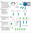
Steps of transgenesis by PB transposon vector. Schematic generation of hSSTR4 transgenic mice (AE). White mice symbolize Sstr4 knockout (KO) mice lacking hSSTR4 transgene, green mice symbolize the correctly inserted hSSTR4 transgene. The transgene copies are named Chr3, Chr10 and ChrX (located in chromosomes 3, 10 and X, respectively) and U1 and U2 (the two copies in an unknown location).
Figure 3
Figure 3
The insertion site of the PB transposon in chromosome 3 of the Sstr4 KO mouse genome, along with the primers used for genotyping. Mouse chromosome 3 showing the location of the transgene insertion in the PB transposase recognition site (TTAA) at the original position of 70,039,120th base pair (*), which was located between the neighboring genes (Otol1 and Sis). Primer sites are shown in the transgene, which integrated in the chromosome 3 used for the first genotyping test of the F0 generation (TR1-4), for verifying the sequencing of the hSSTR4 coding sequence (TR ST4 1–2), for ligation-mediated PCR (LM-V primers) and for present routine genotyping PCR (LM-V3 and Chr3pr2-3, marked with blue).
Figure 4
Figure 4
In vivo bioluminescent imaging of the luciferase reporter protein co-expressed with the hSSTR4 gene. Representative images show the differences of expression pattern and luminescence intensity of luciferase in the different mouse lines (A). Scatter plot with bars show the means ± SEM with the individual data points of the luminescence intensity in equal size areas of the head corresponding to the brain (B). One-way ANOVA, * p < 0.0001, N = 6–19/genotype.
Figure 5
Figure 5
Relative mouse Sstr4 and human SSTR4 mRNA expression levels in wild-type (WT) and humanized (Chr3, U1 and U2) mice, respectively. The diagram shows RT-qPCR results (2−ΔCt) relative to the beta actin (Actb) mRNA reference gene in the tested organs. Cortex, OB and TG stand for cerebral cortex, olfactory bulb and trigeminal ganglion, respectively. Each column shows the mean ± SEM. The significant differences between the WT and Chr3 mice are indicated with an asterisk above the Chr3 column. Kruskal-Wallis test with Dunn’s post-test; * p < 0.01; N = 3–7/genotype.
Figure 6
Figure 6
Expression of the hSSTR4 mRNA in the mouse primary somatosensory cortex. Representative confocal images from Chr3 homozygote mice. Human SSTR4 (red), mouse Vglut1 (green) and mouse Gad1 (white) mRNA expressions counterstained with 4′,6-diamidino-2-phenylindole (DAPI) (blue) were shown in the layer I-VI (A), the layer II-III (B) and the layer V (C) of the primary somatosensory cortex (S1, Bregma −1.46 mm). Scale bar: 50 µm, and inset scale bar: 10 µm.
Figure 7
Figure 7
Expression of the hSSTR4 mRNA in the mouse hippocampus and piriform cortex. Representative confocal images from Chr3 homozygote mice. Human SSTR4 (red), mouse Vglut1 (green) and mouse Gad1 (white) mRNA expressions counterstained with DAPI (blue) were shown in the CA1-CA2 region (Bregma −1.46 mm, (A)), CA1 region of the hippocampus (Bregma −1.46 mm, (B)) and the piriform cortex (Pir, Bregma −1.46 mm, (C)). Scale bar: 50 µm, and inset scale bar: 10 µm.
Figure 8
Figure 8
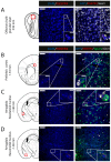
Expression of the hSSTR4 mRNA in the mouse olfactory bulb, prelimbic cortex and amygdala. Representative confocal images from Chr3 homozygote mice. Human SSTR4 (red), mouse Vglut1 (green) and mouse Gad1 (white) mRNA expressions counterstained with DAPI (blue) were shown in the granular layer of the olfactory bulb (OB, Bregma +3.92 mm, (A)), prelimbic cortex (PrL, Bregma +1.54 mm, (B)), basolateral (BLA, (C)) and basomedial (BMA, (D)) nucleus of the amygdala (Bregma −1.46 mm). Scale bar: 50 µm, and inset scale bar: 10 µm.

Similar articles

Cited by

References

    1. Baraban S.C., Tallent M.K. Interneuron Diversity Series: Interneuronal Neuropeptides—Endogenous Regulators of Neuronal Excitability. Trends Neurosci. 2004;27:135–142. doi: 10.1016/j.tins.2004.01.008. - DOI - PubMed
    1. Helyes Z., Pinter E., Sandor K., Elekes K., Banvolgyi A., Keszthelyi D., Szoke E., Toth D.M., Sandor Z., Kereskai L., et al. Impaired Defense Mechanism against Inflammation, Hyperalgesia, and Airway Hyperreactivity in Somatostatin 4 Receptor Gene-Deleted Mice. Proc. Natl. Acad. Sci. USA. 2009;106:13088–13093. doi: 10.1073/pnas.0900681106. - DOI - PMC - PubMed
    1. Sándor K., Elekes K., Szabó Á., Pintér E., Engström M., Wurster S., Szolcsányi J., Helyes Z. Analgesic Effects of the Somatostatin Sst4 Receptor Selective Agonist J-2156 in Acute and Chronic Pain Models. Eur. J. Pharmacol. 2006;539:71–75. doi: 10.1016/j.ejphar.2006.03.082. - DOI - PubMed
    1. Pintér E., Pozsgai G., Hajna Z., Helyes Z., Szolcsányi J. Neuropeptide Receptors as Potential Drug Targets in the Treatment of Inflammatory Conditions. Br. J. Clin. Pharmacol. 2014;77:5–20. doi: 10.1111/bcp.12097. - DOI - PMC - PubMed
    1. Matsuoka N., Maeda N., Yamaguchi I., Satoh M. Possible Involvement of Brain Somatostatin in the Memory Formation of Rats and the Cognitive Enhancing Action of FR121196 in Passive Avoidance Task. Brain Res. 1994;642:11–19. doi: 10.1016/0006-8993(94)90900-8. - DOI - PubMed

Substances