

Lipofilling in Breast Oncological Surgery: A Safe Opportunity or Risk for Cancer Recurrence?
- PMID: 33916703
- PMCID: PMC8038405
- DOI: 10.3390/ijms22073737
Lipofilling in Breast Oncological Surgery: A Safe Opportunity or Risk for Cancer Recurrence?
Abstract
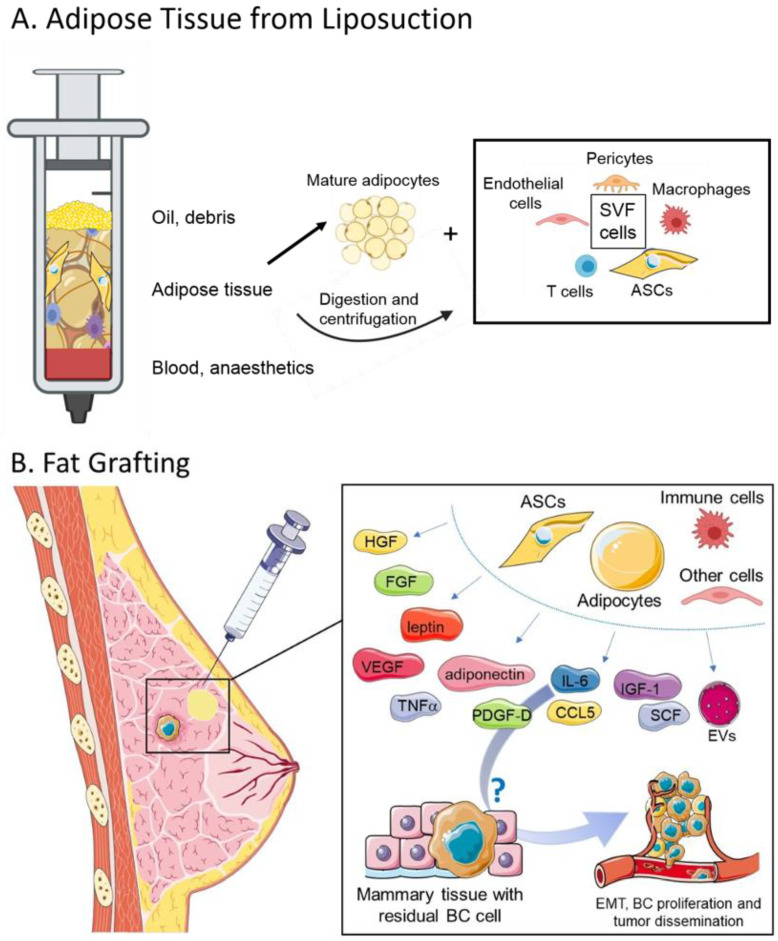
Lipofilling (LF) is a largely employed technique in reconstructive and esthetic breast surgery. Over the years, it has demonstrated to be extremely useful for treatment of soft tissue defects after demolitive or conservative breast cancer surgery and different procedures have been developed to improve the survival of transplanted fat graft. The regenerative potential of LF is attributed to the multipotent stem cells found in large quantity in adipose tissue. However, a growing body of pre-clinical evidence shows that adipocytes and adipose-derived stromal cells may have pro-tumorigenic potential. Despite no clear indication from clinical studies has demonstrated an increased risk of cancer recurrence upon LF, these observations challenge the oncologic safety of the procedure. This review aims to provide an updated overview of both the clinical and the pre-clinical indications to the suitability and safety of LF in breast oncological surgery. Cellular and molecular players in the crosstalk between adipose tissue and cancer are described, and heterogeneous contradictory results are discussed, highlighting that important issues still remain to be solved to get a clear understanding of LF safety in breast cancer patients.
Keywords: adipose tissue; adipose-derived stromal cells; breast cancer; breast cancer recurrence; breast reconstruction; lipofilling; oncological safety; pre-clinical studies.
Conflict of interest statement
The authors declare no conflict of interest.
Figures
References
-
- Zhang Y., Daquinag A., Traktuev D.O., Amaya-Manzanares F., Simmons P.J., March K.L., Pasqualini R., Arap W., Kolonin M.G. White adipose tissue cells are recruited by experimental tumors and promote cancer progression in mouse models. Cancer Res. 2009;69:5259–5266. doi: 10.1158/0008-5472.CAN-08-3444. - DOI - PMC - PubMed
Publication types
MeSH terms
LinkOut - more resources
Full Text Sources
Other Literature Sources
Medical
