Review
. 2021 Apr 3;7(4):273.
doi: 10.3390/jof7040273.

Mycotic Keratitis-A Global Threat from the Filamentous Fungi

Affiliations
Review

Mycotic Keratitis-A Global Threat from the Filamentous Fungi

Jeremy J Hoffman et al. J Fungi (Basel). .

Abstract

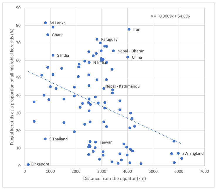
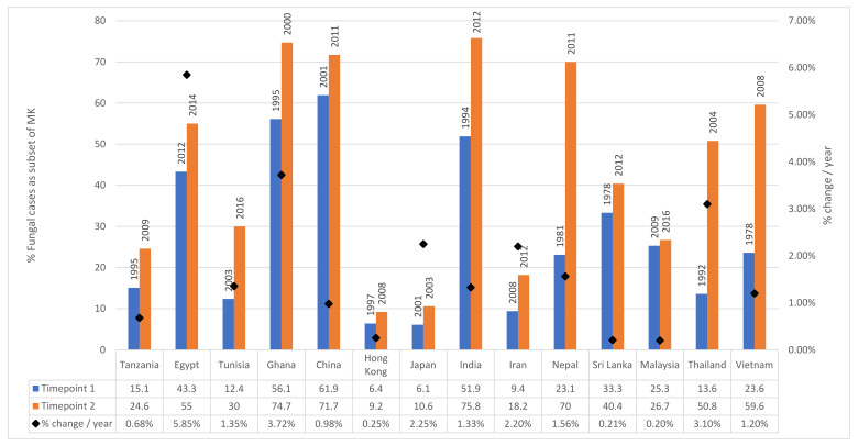
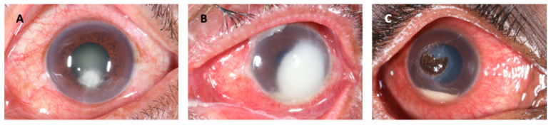
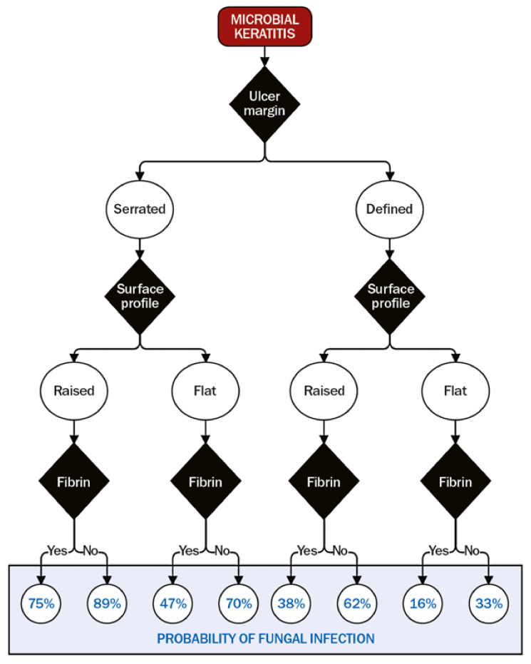
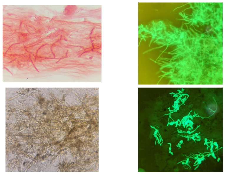
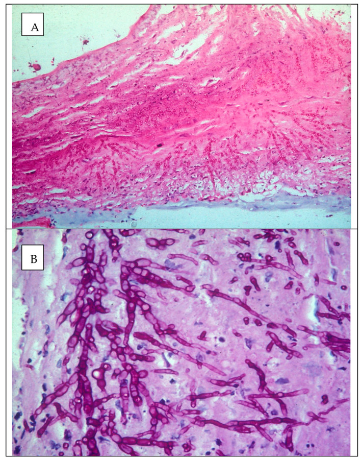
Mycotic or fungal keratitis (FK) is a sight-threatening disease, caused by infection of the cornea by filamentous fungi or yeasts. In tropical, low and middle-income countries, it accounts for the majority of cases of microbial keratitis (MK). Filamentous fungi, in particular Fusarium spp., the aspergilli and dematiaceous fungi, are responsible for the greatest burden of disease. The predominant risk factor for filamentous fungal keratitis is trauma, typically with organic, plant-based material. In developed countries, contact lens wear and related products are frequently implicated as risk factors, and have been linked to global outbreaks of Fusarium keratitis in the recent past. In 2020, the incidence of FK was estimated to be over 1 million cases per year, and there is significant geographical variation; accounting for less than 1% of cases of MK in some European countries to over 80% in parts of south and south-east Asia. The proportion of MK cases is inversely correlated to distance from the equator and there is emerging evidence that the incidence of FK may be increasing. Diagnosing FK is challenging; accurate diagnosis relies on reliable microscopy and culture, aided by adjunctive tools such as in vivo confocal microscopy or PCR. Unfortunately, these facilities are infrequently available in areas most in need. Current topical antifungals are not very effective; infections can progress despite prompt treatment. Antifungal drops are often unavailable. When available, natamycin is usually first-line treatment. However, infections may progress to perforation in ~25% of cases. Future work needs to be directed at addressing these challenges and unmet needs. This review discusses the epidemiology, clinical features, diagnosis, management and aetiology of FK.

Keywords: Aspergillus; Fusarium; blindness; dematiaceous fungi; epidemiology; fungal keratitis; microbial keratitis; microbiology; mycotic keratitis.


Conflict of interest statement

The authors declare no conflict of interest. The funders had no role in the design of the study; in the collection, analyses, or interpretation of data; in the writing of the manuscript, or in the decision to publish the results.

Figures

Figure 1
Figure 1
Fungal keratitis in a patient presenting to an ophthalmic hospital in Nepal. The causative organism was confirmed to be Fusarium sp. on culture. (A): The conjunctiva is hyperaemic, causing the eye to be red. There is a white corneal infiltrate with feathery serrated margins and satellite lesions present. There is also a small hypopyon. (B): The same eye as viewed with a cobalt blue filter after instillation of topical fluorescein. The area staining in green represents a defect in the corneal epithelium.
Figure 2
Figure 2
The progression of a patient with fungal keratitis caused by Aspergillus sp. This patient presented early in the course of the disease with a relatively small corneal ulcer, with serrated feathery margins to the corneal infiltrate (A). Despite intense, appropriate, prompt treatment with topical natamycin 5%, the corneal infiltrate increased in size, ultimately perforating, and was temporarily treated with corneal gluing and bandage contact lens insertion (B). The patient ultimately underwent a therapeutic penetrating keratoplasty (C).
Figure 3
Figure 3
Fungal keratitis as a proportion of all culture positive microbial keratitis cases, by distance from the equator, with select locations shown, with calculated line of best fit given (dotted line, y = − 0.0069x + 54.696).
Figure 4
Figure 4
Percentage of fungal cases as a subset of MK plotted by country at two timepoints. Timepoint 1 represents the earliest year for which values were available, Timepoint 2 represents the latest year for which values are available. The years for the two studies are given as labels. The percentage change per year (calculated from the difference between the two timepoints) is plotted against the secondary y-axis.
Figure 5
Figure 5
Differing clinical phenotypes of filamentous fungal keratitis depending on the fungal organism. (A): Fusarium sp. Note the serrated or feathery margins, satellite lesions, non-yellow infiltrate and lack of hypopyon. (B): Aspergillus sp. Note less obviously serrated margins compared to (A), raised profile, hypopyon. (C): Curvularia sp. Note the raised, pigmented infiltrate, in addition to the hypopyon.
Figure 6
Figure 6
Algorithm for determining the probability of fungal keratitis [163]. The black diamonds are decision points about three clinical features: ulcer/infiltrate margin, surface profile, and anterior chamber fibrin. These probabilities are based on data presented in Thomas et al. [2]. This is reproduced here from [163] with permission under a CC BY-NC 4.0 license (https://creativecommons.org/licenses/by-nc/4.0/, accessed on 16 March 2021).
Figure 7
Figure 7
Microscopic appearance of filamentous fungal hyphae in corneal tissue (corneal scrape specimens) using different staining techniques. Clockwise from top-left: Fungal hyphae in Gram-stained corneal smear (magnification 1000x, oil immersion); fungal hyphae visible with CFW, Curvularia sp. stained with CFW, pigmented hyphae (Curvularia sp.) in a KOH preparation (magnification 400x). These images were taken using an afocal photography technique; the camera zoom was used for additional magnification.
Figure 8
Figure 8
In vivo confocal microscopy of fungal keratitis (A) clinical image; (B) In vivo confocal microscopy scan of the same cornea showing extensive, branching fungal hyphae. Scale bar 100 μm. (C) Light microscopy demonstrated septate fungal hyphae, visible on Gram staining (magnification 1000x, oil immersion); (D) and KOH preparation (magnification 400x). Images (C,D) were taken using an afocal photography technique; the camera zoom was used for additional magnification.
Figure 9
Figure 9
Algorithm for diagnosing fungal keratitis.
Figure 10
Figure 10
Examples of dematiaceous fungal genera isolated from cases of fungal keratitis stained with LPCB. Clockwise from top-left: Curvularia sp., Bipolaris sp. (magnification 400x); Alternaria sp., Exserohilum sp. (magnification x1000, oil immersion). These images were taken using an afocal photography technique; the camera zoom was used for additional magnification.
Figure 11
Figure 11
Histology section of a corneal button infected with Scedosporium apiospermum stained with H&E/PAS (A—magnification ×100, B—magnification ×1000, oil immersion).

Similar articles

Cited by

References

    1. Burton M.J., Pithuwa J., Okello E., Afwamba I., Onyango J.J., Oates F., Chevallier C., Hall A.B. Microbial Keratitis in East Africa: Why are the Outcomes so Poor? Ophthalmic Epidemiol. 2011;18:158–163. doi: 10.3109/09286586.2011.595041. - DOI - PMC - PubMed
    1. Thomas P.A., Leck A.K., Myatt M. Characteristic clinical features as an aid to the diagnosis of suppurative keratitis caused by filamentous fungi. Br. J. Ophthalmol. 2005;89:1554–1558. doi: 10.1136/bjo.2005.076315. - DOI - PMC - PubMed
    1. Brown L., Leck A.K., Gichangi M., Burton M.J., Denning D.W. The global incidence and diagnosis of fungal keratitis. Lancet Infect. Dis. 2020 doi: 10.1016/S1473-3099(20)30448-5. - DOI - PubMed
    1. Srinivasan M., Gonzales C.A., George C., Cevallos V., Mascarenhas J.M., Asokan B., Wilkins J., Smolin G., Whitcher J.P. Epidemiology and aetiological diagnosis of corneal ulceration in Madurai, south India. Br. J. Ophthalmol. 1997;81:965–971. doi: 10.1136/bjo.81.11.965. - DOI - PMC - PubMed
    1. Whitcher J.P., Srinivasan M., Upadhyay M.P. Corneal blindness: A global perspective. Bull. World Health Organ. 2001;79:214–221. - PMC - PubMed

LinkOut - more resources