Review
. 2021 Apr 7;13(4):629.
doi: 10.3390/v13040629.

Rhinovirus and Cell Death

Affiliations
Review

Rhinovirus and Cell Death

Shannic-Le Kerr et al. Viruses. .

Abstract

Rhinoviruses (RVs) are the etiological agents of upper respiratory tract infections, particularly the common cold. Infections in the lower respiratory tract is shown to cause severe disease and exacerbations in asthma and COPD patients. Viruses being obligate parasites, hijack host cell pathways such as programmed cell death to suppress host antiviral responses and prolong viral replication and propagation. RVs are non-enveloped positive sense RNA viruses with a lifecycle fully contained within the cytoplasm. Despite decades of study, the details of how RVs exit the infected cell are still unclear. There are some diverse studies that suggest a possible role for programmed cell death. In this review, we aimed to consolidate current literature on the impact of RVs on cell death to inform future research on the topic. We searched peer reviewed English language literature in the past 21 years for studies on the interaction with and modulation of cell death pathways by RVs, placing it in the context of the broader knowledge of these interconnected pathways from other systems. Our review strongly suggests a role for necroptosis and/or autophagy in RV release, with the caveat that all the literature is based on RV-A and RV-B strains, with no studies to date examining the interaction of RV-C strains with cell death pathways.

Keywords: apoptosis; autophagy; cell death pathways; lifecycle; necroptosis; necrosis; rhinovirus.


Conflict of interest statement

The authors declare no conflict of interest.

Figures

Figure 1
Figure 1
Rhinovirus lifecycle. (1) Binding of the virus to receptors on the plasma membrane initiates (2) endocytosis. The intake of the virion is followed by (3) uncoating of the capsid and release of the (+)ss-RNA into the host cytoplasm, which is (4) translated and processed into structural and non-structural proteins. The viral RNA dependent RNA polymerase converts the viral genome into (−)ss-RNA, that is (5) replicated into new (+)ss-RNA genomes. The new genomes become the template for translation into new viral proteins and replication into new genomes. The final stages involve (6) assembly of the structural proteins and RNA genome into capsids and (7) release of new infectious virions via lysis or non-lytic mechanisms. Adapted from [5,13,16,28]. Created with BioRender.com, accessed on 12 December 2020.
Figure 2
Figure 2
Extrinsic and Intrinsic apoptosis. (A) Extrinsic apoptosis. Ligand binding results in activation of cytoplasmic adapter proteins and recruitment of death domains on FasL/FasR or TNF-α/TNFR1. This activates FADD, which can directly activate pro-caspase-8 or can form a complex with TRADD and RIP, bind to TNF-α/TNFR1 and then induce auto-catalytic activation of procaspase-8. Activated caspase 8 initiates the execution phase of apoptosis. (B) Intrinsic apoptosis. Initiated by non-receptor-mediated stimuli that generate mitochondrial-initiated intracellular signals. These signals initiate a cascade of events resulting in the formation of an apoptosome. The apoptosome activates the executioner caspases-3/6/7 and initiates apoptosis. Adapted from [5,46,47]. Created with BioRender.com, accessed on 12 December 2020.
Figure 3
Figure 3
Three autophagy pathways (a) Macroautophagy: cytosolic material is transported to the lysosome using double membrane-bound vesicles which engulf the material and then fuse to the lysosome (b) Chaperone mediated autophagy: proteins bound to HSC70 being translocated to the lysosomal membrane to be unfolded and degraded. (c) Microautophagy: cytosolic components being directly taken up by the lysosome. Adapted from [60,61]. Created with BioRender.com, accessed on 25 January 2020.
Figure 4
Figure 4
Necroptopic signaling pathway. TNFa is bound to the extracellular domain of the TNFR1. TNFR1 triggers downstream signaling by forming complex I with TRADD, FADD, RIP1, and several E3 ubiquitination ligases, TRAF2/5 and cIAP1/2. Deubiquitinated RIP1 is transferred from complex I to the cytoplasm and prompts the recruitment of complex Ia. Complex Ia is comprised of TRADD, FADD, RIP1 and caspase 8. Caspase 8 is inhibited in complex Ib, allowing for RIP1 to bind to RIP3. Bound RIP3 recruits and phosphorylates MLKL. Activated MLKL disrupts the membrane leading to permeabilization, swelling and rupturing. Adapted from [51,79,80,81,82]. Created with BioRender.com, accessed on 15 January 2021.
Figure 5
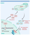
Figure 5
Parthanatos pathway. (a) Severe or prolonged DNA damage, oxidative stress, hypoxia, hypoglycemia or inflammation (b) triggers the activation of the enzyme poly(ADP-ribose) polymerase (PARP). (c) This triggers a series of cytotoxic effects such as NAD+ and ATP depletion and accumulation of poly(ADP-ribosyl)ated proteins (PAR) at the mitochondria. (d) PAR bind to apoptosis-inducing factor (AIF), decrease in mitochondrial membrane potential (MMP) and prompts release of AIF into the cytoplasm. (e) AIF then translocates to the nucleus and mediates large-scale DNA fragmentation and chromatin condensation [91,92,93,96]. Created using BioRender.com, accessed on 31 January 2021.

References

    1. Zhang T., Yin C., Boyd D.F., Quarato G., Ingram J.P., Shubina M., Ragan K.B., Ishizuka T., Crawford J.C., Tummers B., et al. Influenza Virus Z-RNAs Induce ZBP1-Mediated Necroptosis. Cell. 2020;180:1115–1129. doi: 10.1016/j.cell.2020.02.050. - DOI - PMC - PubMed
    1. Kuo C.-Y., Chiu V., Hsieh P.-C., Huang C.-Y., Huang S.J., Tzeng I.S., Tsai F.-M., Chen M.-L., Liu C.-T., Chen Y.-R. Chrysophanol attenuates hepatitis B virus X protein-induced hepatic stellate cell fibrosis by regulating endoplasmic reticulum stress and ferroptosis. J. Pharmacol. Sci. 2020;144:172–182. doi: 10.1016/j.jphs.2020.07.014. - DOI - PubMed
    1. Suwanmanee S., Luplertlop N. Immunopathogenesis of Dengue Virus-Induced Redundant Cell Death: Apoptosis and Pyroptosis. Viral Immunol. 2017;30:13–19. doi: 10.1089/vim.2016.0092. - DOI - PubMed
    1. Kim J.H., Kim J., Roh J., Park C.S., Seoh J.Y., Hwang E.S. Reactive oxygen species-induced parthanatos of immunocytes by human cytomegalovirus-associated substance. Microbiol. Immunol. 2018;62:229–242. doi: 10.1111/1348-0421.12575. - DOI - PMC - PubMed
    1. Croft S.N., Walker E.J., Ghildyal R. Picornaviruses and Apoptosis: Subversion of Cell Death. mBio. 2017;8:e01009-17. doi: 10.1128/mBio.01009-17. - DOI - PMC - PubMed

LinkOut - more resources