. 2021 Apr 7;13(4):632.
doi: 10.3390/v13040632.

Development and Characterization of a cDNA-Launch Recombinant Simian Hemorrhagic Fever Virus Expressing Enhanced Green Fluorescent Protein: ORF 2b' Is Not Required for In Vitro Virus Replication

Affiliations

Development and Characterization of a cDNA-Launch Recombinant Simian Hemorrhagic Fever Virus Expressing Enhanced Green Fluorescent Protein: ORF 2b' Is Not Required for In Vitro Virus Replication

Yingyun Cai et al. Viruses. .

Abstract

Simian hemorrhagic fever virus (SHFV) causes acute, lethal disease in macaques. We developed a single-plasmid cDNA-launch infectious clone of SHFV (rSHFV) and modified the clone to rescue an enhanced green fluorescent protein-expressing rSHFV-eGFP that can be used for rapid and quantitative detection of infection. SHFV has a narrow cell tropism in vitro, with only the grivet MA-104 cell line and a few other grivet cell lines being susceptible to virion entry and permissive to infection. Using rSHFV-eGFP, we demonstrate that one cricetid rodent cell line and three ape cell lines also fully support SHFV replication, whereas 55 human cell lines, 11 bat cell lines, and three rodent cells do not. Interestingly, some human and other mammalian cell lines apparently resistant to SHFV infection are permissive after transfection with the rSHFV-eGFP cDNA-launch plasmid. To further demonstrate the investigative potential of the infectious clone system, we introduced stop codons into eight viral open reading frames (ORFs). This approach suggested that at least one ORF, ORF 2b', is dispensable for SHFV in vitro replication. Our proof-of-principle experiments indicated that rSHFV-eGFP is a useful tool for illuminating the understudied molecular biology of SHFV.

Keywords: Arteriviridae; Nidovirales; SHFV; Simarterivirinae; cell tropism; infectious clone; reverse genetics; simarterivirin; simarterivirus; simian hemorrhagic fever virus.


Conflict of interest statement

The authors declare no conflict of interest.

Figures

Figure 1
Figure 1
Schematics of simian hemorrhagic fever virus (SHFV) cDNA-launch infectious clones. (A) The SHFV genome is a linear, unsegmented, polyadenylated, positive-sense RNA. Open reading frames (ORFs) are shown as colored rectangles. ORF1a is translated directly from the genomic RNA into polyprotein 1a (pp1a). A -2 programmed ribosomal frameshift results in nonstructural protein 2 transframe (nsp2TF), whereas a -1 frameshift fuses ORF1a and ORF1b, resulting in polyprotein 1ab (pp1b). pp1a and pp1ab are co- and post-translationally cleaved to yield numerous SHFV nonstructural proteins (nsps). The remaining ORFs encode the SHFV structural proteins, which are translated from a nested set of subgenomic RNAs. E, envelope protein; GP, glycoprotein; M, matrix protein; N, nucleocapsid protein. (B) Constructed plasmid pCMV-SHFV, encoding wild-type SHFV. (C) Constructed plasmid pCMV-SHFV-eGFP, encoding SHFV-eGFP.
Figure 2
Figure 2
Growth kinetics of wild-type simian hemorrhagic fever virus variant NIH LVR42-0/M6941 (SHFV), recombinant wild-type SHFV (rSHFV), and rSHFV expressing enhanced green fluorescent protein (rSHFV-eGFP). (A) MA-104 cells were mock-exposed (control), exposed to SHFV (multiplicity of infection (MOI) of 0.1), transfected with pCMV-SHFV, or transfected with pCMV-SHFV-eGFP. Top row: Microscopic images reveal the cytopathic effect caused by SHFV, rSHFV, and rSHFV-eGFP infection at 16 h post-exposure/transfection. Middle row: Expression of eGFP was observed using epifluorescent microscopy. Bottom row: plaque morphologies of the three viruses. (B) MA-104 cells were exposed to all three viruses at MOIs of 0.1 (left) and 1 (right), tissue culture supernatants were harvested at the indicated time points post-exposure, and virus titers were determined by plaque assay. Graphs of viral titers represent the means ± the standard deviations of triplicate samples from one of two independent experiments.
Figure 3
Figure 3
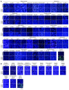
Cellular tropism of simian hemorrhagic fever virus (SHFV): Recombinant wild-type SHFV expressing enhanced green fluorescent protein-expressing (rSHFV-eGFP) does not infect human or bat cells. (A) 53 adherent cells of the human NCI-60 cancer cell line panel and (B) 11 bat cell lines were exposed to rSHFV-eGFP at a multiplicity of infection of 3. Shown are high-content images of mock-exposed controls and virus-exposed cells counterstained with Hoechst33342 at 72 h or 24 h post-exposure for positive control MA-104 cells.
Figure 4
Figure 4
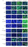
Cellular tropism of simian hemorrhagic fever virus (SHFV): Recombinant wild-type SHFV expressing enhanced green fluorescent protein-expressing (rSHFV-eGFP) infects hispid cotton rat lung (CRL) cells. (A) Grivet cells, (B) human cells that are not part of the human NCI-60 cancer cell line panel, and (C) rodent cells were exposed to rSHFV-eGFP at a multiplicity of infection of 3. Tissue culture supernatants were harvested at 72 h post-exposure, cells were counterstained with Hoechst 33342, and high-content images of exposed cells or mock-infected cells were taken. Supernatants of virus-exposed cells were also subjected to plaque assay; obtained titers are printed beneath the images. Uninfected cells were also transfected with pCMV-SHFV-eGFP or a control plasmid expressing eGFP only. At 48 h post-transfection, cells were counterstained with Hoechst 33342, high-content images were taken, supernatants were harvested, and viral titers were measured by plaque assay; obtained titers are indicated beneath the images.
Figure 5
Figure 5
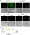
Cellular tropism of simian hemorrhagic fever virus (SHFV): Recombinant wild-type SHFV expressing enhanced green fluorescent protein-expressing (rSHFV-eGFP) infects hispid cotton rat lung (CRL) cells, western gorilla RPGor53 primary fibroblasts, and chimpanzee S008397 and RP00226 primary fibroblasts. (A) CRL cells, western gorilla primary fibroblasts, and common chimpanzee primary fibroblasts were exposed to rSHFV-eGFP at a multiplicity of infection (MOI) of 3. eGFP expression and cytopathic effect were observed at 24 h post-exposure. (B) Cells were exposed to wild-type SHFV at an MOI of 3. Tissue culture supernatants were harvested at the indicated time points post-exposure, and virus titers were determined by plaque assay. Graphs of viral titers represent the means ± the standard deviations of triplicate samples from one of two independent experiments.
Figure 6
Figure 6
Genetic stability of recombinant wild-type simian hemorrhagic fever virus (SHFV) expressing enhanced green fluorescent protein-expressing (rSHFV-eGFP) in MA-104 cells. rSHFV-eGFP was serially passaged on MA-104 cells. Cells were exposed to rSHFV-eGFP at a multiplicity of infection (MOI) of 1, tissue culture supernatants were collected (Passage 1 [p1]) at 48 h post-exposure, and virus titers were determined by plaque assay. Fresh MA-104 cells were infected with tissue culture supernatants at an MOI of 1. This process was serially repeated through Passage 5 (p5). eGFP expression (green) was measured by epifluorescent microscopy (top row). Cells were imaged by compound microscopy (bottom row).
Figure 7
Figure 7
Minor structural protein E’ is dispensable for infectious simian hemorrhagic fever virus (SHFV) particle production in vitro. (A) To analyze the functional importance of SHFV minor structural proteins, stop codons (red) were introduced into the respective open reading frames (ORFs) of pCMV-SHFV-eGFP without altering the amino acids encoded in overlapping ORFs. (B) MA-104 cells were transfected with the pCMV-SHFV-eGFP mutants. Supernatants were harvested at 48 h p.t. and subjected to plaque assay. Top row: epifluorescent microscopic images of eGFP-generated fluorescence. Bottom row: Compound microscopic images of cells with plaque-assay titers printed beneath them. (C) MA-104 cells were exposed to supernatants harvested in (B) and eGFP expression was measured by epifluorescence microscopy.
Figure 7
Figure 7
Minor structural protein E’ is dispensable for infectious simian hemorrhagic fever virus (SHFV) particle production in vitro. (A) To analyze the functional importance of SHFV minor structural proteins, stop codons (red) were introduced into the respective open reading frames (ORFs) of pCMV-SHFV-eGFP without altering the amino acids encoded in overlapping ORFs. (B) MA-104 cells were transfected with the pCMV-SHFV-eGFP mutants. Supernatants were harvested at 48 h p.t. and subjected to plaque assay. Top row: epifluorescent microscopic images of eGFP-generated fluorescence. Bottom row: Compound microscopic images of cells with plaque-assay titers printed beneath them. (C) MA-104 cells were exposed to supernatants harvested in (B) and eGFP expression was measured by epifluorescence microscopy.
Figure 8
Figure 8
Replacement of the recombinant wild-type simian hemorrhagic fever virus (SHFV) expressing enhanced green fluorescent protein-expressing (rSHFV-eGFP) open reading frames (ORFs) encoding the minor structural proteins E, GP2, GP3, and GP4 with ORFs encoding presumed EAV orthologs does not expand SHFV cell tropism. MA-104 cells were transfected with a plasmid encoding rSHFV-eGFP control (top) or chimeric rSHFV-eGFP (bottom). At 48 h post-transfection, eGFP expression was measured by epifluorescence microscopy. Supernatants (containing rSHFV-eGFP or rSHFV-eGFP-EAV-ORF2ab34) were harvested and used to expose fresh MA-104, Vero, and BHK-21 cells at a multiplicity of infection of 1. Success of infection was measured by epifluorescent microscopy.
Figure 9
Figure 9
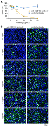
CD163 is a crucial simian hemorrhagic fever virus (SHFV) cell entry factor. (A) MA-104 cells were incubated with different concentrations of goat anti-human CD163 antibody or control goat immunoglobulin G (IgG) at 37 °C for 1 h. The treated cells were exposed to recombinant wild-type SHFV expressing enhanced green fluorescent protein-expressing (rSHFV-eGFP) at a multiplicity of infection of 5 at 37 °C for 1 h in the presence of antibodies. The percentage of rSHFV-eGFP-infected cells in the presence of increasing goat anti-CD163 antibody (yellow) and control goat IgG control (blue) is indicated at 48 h post-exposure. Error bars indicate the standard deviation of triplicate samples from one of two independent experiments. (B) Representative images of the experiment described in (A).

Similar articles

Cited by

References

    1. Шевцoва З.В., Куксoва М.И., Джикидзе Э.К., Крылoва Р.И., Данькo Л.В. Экспериментальнoе изучение гемoррагическoй лихoрадки oбезьян. In: Lapin B.A., editor. Biology and Pathology of Monkeys, Studies of Human Diseases in Experiments on Monkeys. Materials of Symposium in Sukhumi, 17–22 October 1966. Academy of Medical Sciences of the U.S.S.R., Institute of Experimental Pathology and Therapy; Tbilisi, Georgian Soviet Socialist Republic, USSR: 1966. pp. 146–150.
    1. Шевцoва З.В. Изучение этиoлoгии гемoррагическoй лихoрадки oбезьян. Вoпр. Вирусoл. 1967;12:47–51. - PubMed
    1. Palmer A.E., Allen A.M., Tauraso N.M., Shelokov A. Simian hemorrhagic fever. I. Clinical and epizootiologic aspects of an outbreak among quarantined monkeys. Am. J. Trop. Med. Hyg. 1968;17:404–412. doi: 10.4269/ajtmh.1968.17.404. - DOI - PubMed
    1. Lauck M., Alkhovsky S.V., Bào Y., Bailey A.L., Shevtsova Z.V., Shchetinin A.M., Vishnevskaya T.V., Lackemeyer M.G., Postnikova E., Mazur S., et al. Historical outbreaks of simian hemorrhagic fever in captive macaques were caused by distinct arteriviruses. J. Virol. 2015;89:8082–8087. doi: 10.1128/JVI.01046-15. - DOI - PMC - PubMed
    1. Tauraso N.M., Shelokov A., Palmer A.E., Allen A.M. Simian hemorrhagic fever. III. Isolation and characterization of a viral agent. Am. J. Trop. Med. Hyg. 1968;17:422–431. doi: 10.4269/ajtmh.1968.17.422. - DOI - PubMed

Publication types

LinkOut - more resources