

A Review of the Role of Endo/Sarcoplasmic Reticulum-Mitochondria Ca2+ Transport in Diseases and Skeletal Muscle Function
- PMID: 33917091
- PMCID: PMC8067840
- DOI: 10.3390/ijerph18083874
A Review of the Role of Endo/Sarcoplasmic Reticulum-Mitochondria Ca2+ Transport in Diseases and Skeletal Muscle Function
Abstract
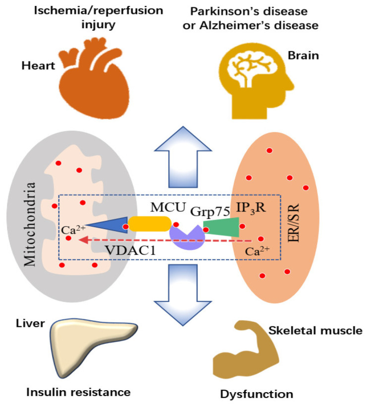
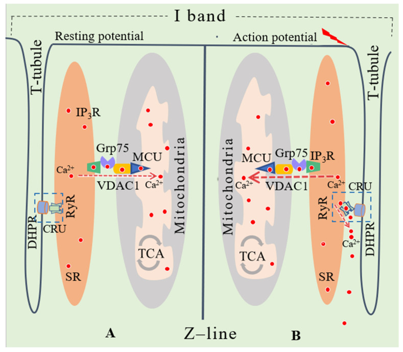
The physical contact site between a mitochondrion and endoplasmic reticulum (ER), named the mitochondria-associated membrane (MAM), has emerged as a fundamental platform for regulating the functions of the two organelles and several cellular processes. This includes Ca2+ transport from the ER to mitochondria, mitochondrial dynamics, autophagy, apoptosis signalling, ER stress signalling, redox reaction, and membrane structure maintenance. Consequently, the MAM is suggested to be involved in, and as a possible therapeutic target for, some common diseases and impairment in skeletal muscle function, such as insulin resistance and diabetes, obesity, neurodegenerative diseases, Duchenne muscular dystrophy, age-related muscle atrophy, and exercise-induced muscle damage. In the past decade, evidence suggests that alterations in Ca2+ transport from the ER to mitochondria, mediated by the macromolecular complex formed by IP3R, Grp75, and VDAC1, may be a universal mechanism for how ER-mitochondria cross-talk is involved in different physiological/pathological conditions mentioned above. A better understanding of the ER (or sarcoplasmic reticulum in muscle)-mitochondria Ca2+ transport system may provide a new perspective for exploring the mechanism of how the MAM is involved in the pathology of diseases and skeletal muscle dysfunction. This review provides a summary of recent research findings in this area.
Keywords: endo/sarcoplasmic reticulum-mitochondria Ca2+ transport; mitochondria-associated membrane; mitochondrial calcium overload; skeletal muscle function.
Conflict of interest statement
The authors declare no conflict of interest.
Figures
References
-
- Xu H., Guan N., Ren Y.-L., Wei Q.-J., Tao Y.-H., Yang G.-S., Liu X.-Y., Bu D.-F., Zhang Y., Zhu S.-N. IP3R-Grp75-VDAC1-MCU calcium regulation axis antagonists protect podocytes from apoptosis and decrease proteinuria in an Adriamycin nephropathy rat model. BMC Nephrol. 2018;19:140. doi: 10.1186/s12882-018-0940-3. - DOI - PMC - PubMed
-
- Gomez L., Thiebaut P.A., Paillard M., Ducreux S., Abrial M., Crola Da Silva C., Durand A., Alam M.R., Van Coppenolle F., Sheu S.S., et al. The SR/ER-mitochondria calcium crosstalk is regulated by GSK3β during reperfusion injury. Cell Death Differ. 2015;22:1890. doi: 10.1038/cdd.2015.118. - DOI - PMC - PubMed
Publication types
MeSH terms
Substances
LinkOut - more resources
Full Text Sources
Other Literature Sources
Miscellaneous
