Review
. 2021 Apr 7;22(8):3827.
doi: 10.3390/ijms22083827.

Human Mitoribosome Biogenesis and Its Emerging Links to Disease

Affiliations
Review

Human Mitoribosome Biogenesis and Its Emerging Links to Disease

Maria Isabel G Lopez Sanchez et al. Int J Mol Sci. .

Abstract

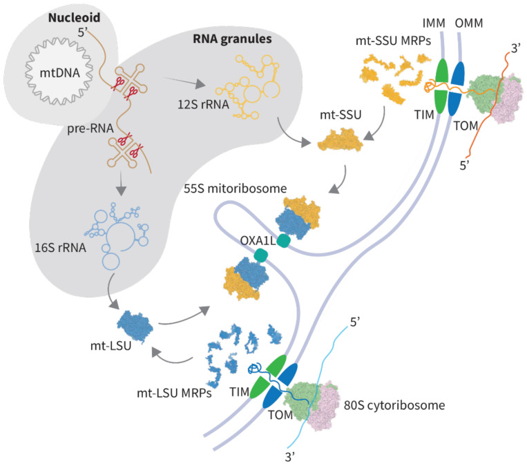
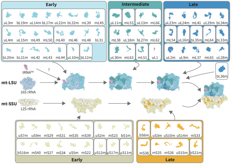
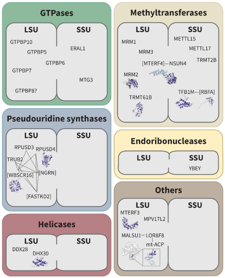
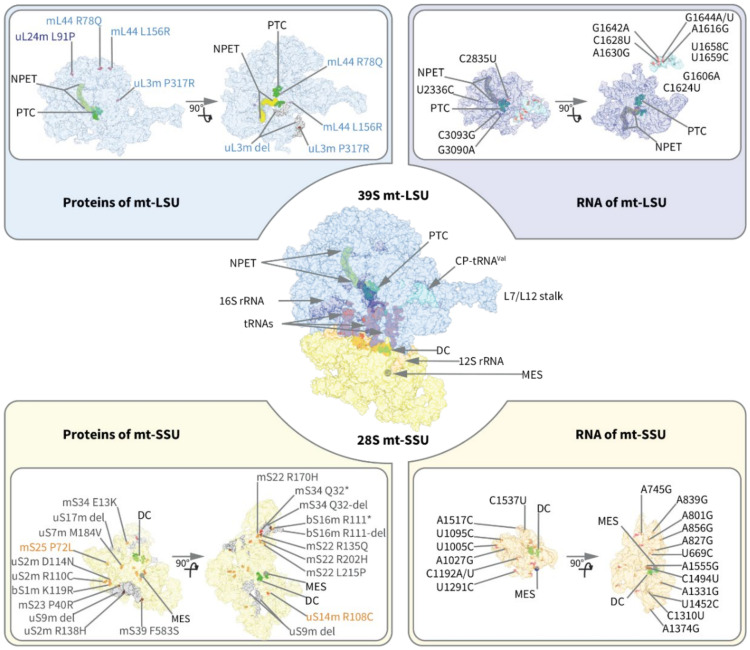
Mammalian mitochondrial ribosomes (mitoribosomes) synthesize a small subset of proteins, which are essential components of the oxidative phosphorylation machinery. Therefore, their function is of fundamental importance to cellular metabolism. The assembly of mitoribosomes is a complex process that progresses through numerous maturation and protein-binding events coordinated by the actions of several assembly factors. Dysregulation of mitoribosome production is increasingly recognized as a contributor to metabolic and neurodegenerative diseases. In recent years, mutations in multiple components of the mitoribosome assembly machinery have been associated with a range of human pathologies, highlighting their importance to cell function and health. Here, we provide a review of our current understanding of mitoribosome biogenesis, highlighting the key factors involved in this process and the growing number of mutations in genes encoding mitoribosomal RNAs, proteins, and assembly factors that lead to human disease.

Keywords: assembly factors; mitochondria; mitochondrial disease; mitoribosome; rRNA.


Conflict of interest statement

The authors declare no conflict of interest.

Figures

Figure 1
Figure 1
Biogenesis of the mitoribosome. The mitoribosome is composed of mitoribosomal proteins (MRPs) and RNAs, which are produced in the cytosol and mitochondria, respectively. The RNA components are derived from the transcription of mtDNA in nucleoids, followed by the RNA maturation in RNA granules. MRPs are synthesized by the 80S cytosolic ribosome and imported into the mitochondrial matrix through the mitochondrial import machinery (TOM and TIM proteins). Next, MRPs and rRNAs are assembled into the mitochondrial large subunit (mt-LSU) and the mitochondrial small subunit (mt-SSU). The complete 55S monosome is anchored to the inner mitochondrial membrane (IMM) and interacts with the translocase OXA1L. OMM-outer mitochondrial membrane.
Figure 2
Figure 2
The assembly pathways of mt-LSU and mt-SSU. Components of the mt-LSU and mt-SSU are assembled in a hierarchical manner. The mt-LSU is assembled from 52 proteins, 16S rRNA, and mt-tRNAVal, whereas the mt-SSU is assembled from 30 proteins and 12S rRNA. Three main stages (early, intermediate, and late stages) have been identified in the mt-LSU, and two main stages (early and late stages) in the mt-SSU assembly pathways. Protein components integrating into the assembly intermediates in clusters are shown together, while individually recruited proteins are shown alone in boxes. Except for bL36m, the incorporation data are based on [16]. bL36m was integrated according to [17]. The timing of mt-tRNAVal incorporation is still unknown (marked by a question mark), as well as the incorporation of mL52, mS37, and bS18m. All structures are based on the model PDB 6ZSG [18]. uL1m is not included in the figure due to its absence in 6ZSG; for uL12m, only the N-terminal structure of uL12m is depicted as it is the only part resolved in the 6ZSG model.
Figure 3
Figure 3
Mitoribosome assembly factors. Mitoribosomal assembly factors and their functional partners. Assembly factors without known enzymatic activity are termed “others”. Certain assembly factors are known to work in protein modules and are thus represented together and connected by dashed lines (NSUN4-MTERF4 [24,25,26]/TFB1M-RBFA [27]/RPUSD3-RPUSD4-TRUB2-WBSCR16-NGRN-FASTKD2 [28,29]). Factors of protein modules, which do not possess the corresponding enzymatic activity, are indicated in squared brackets. Even though RPUSD3 and TRUB2 are pseudouridine synthases, their activity toward 16S rRNA could not be demonstrated so far. GTPBP8 is speculated to participate in the assembly process due to its homology with the bacterial assembly factor. For all other proteins, some experimental evidence exists for their involvement in mitoribosome biogenesis. Available structures are depicted (RPUSD4 (pdb 5UBA), WBSCR16 (pdb 5XGS), DHX30 (pdb 2DB2), MRM2 (pdb 2NYU), TRMT61B (pdb 2B25), TFB1M (pdb 6AAX), RBFA (pdb 2E7G), NSUN4-MTERF4 (pdb 4FP9), and MTERF3 (pdb 3M66)). In the context of the mitoribosome, there is only structural data for the MALSU1 module available (MALSU1-LOR8F8-mt-ACP (PDB 500M)).
Figure 4
Figure 4
Disease-related mutations mapped on the structure of the human mitoribosome. Human mitochondrial ribosome (central panel). The mt-LSU contains mitoribosomal proteins (light blue), 16S mt-rRNA (blue), and CP-tRNAVal (cyan), whereas the mt-SSU includes mitoribosomal proteins (yellow) and 12S mt-rRNA (orange). A-, P-, and E-site tRNA molecules (light red) bind both subunits of the mitoribosome. Key active sites of the mitoribosome: peptidyl transferase center (PTC) and decoding center (DC) are colored in green. Nascent peptide exit tunnel (NPET) is shown on the mt-LSU in yellow. mRNA entry site (MES) is shown on the mt-SSU in blue. Point mutations (red) or deletions (gray) may occur in both LSU (top) and SSU (bottom), targeting either protein components (left-hand side panel) or RNA (right-hand side panel). The colors of the protein names reflect the assembly stage they are incorporated into mitoribosome, according to the kinetic model (see Figure 2 for details). Each component (proteins or RNA) is shown in two orientations, the same as the original 55S monosome and rotated by 90° to depict the intersubunit surface. “del” indicates a complete deletion of a protein, while “X-del” notation indicates that residue X becomes a stop-codon and remaining C-terminal part is not present in the protein. Some of the mutations were not mapped since they are not resolved in the structure (see Table 1 for details).

Similar articles

  • The Diseased Mitoribosome.
    Ferrari A, Del'Olio S, Barrientos A. Ferrari A, et al. FEBS Lett. 2021 Apr;595(8):1025-1061. doi: 10.1002/1873-3468.14024. Epub 2020 Dec 22. FEBS Lett. 2021. PMID: 33314036 Free PMC article. Review.
  • A roadmap for ribosome assembly in human mitochondria.
    Lavdovskaia E, Hanitsch E, Linden A, Pašen M, Challa V, Horokhovskyi Y, Roetschke HP, Nadler F, Welp L, Steube E, Heinrichs M, Mai MM, Urlaub H, Liepe J, Richter-Dennerlein R. Lavdovskaia E, et al. Nat Struct Mol Biol. 2024 Dec;31(12):1898-1908. doi: 10.1038/s41594-024-01356-w. Epub 2024 Jul 11. Nat Struct Mol Biol. 2024. PMID: 38992089 Free PMC article.
  • The human Obg protein GTPBP10 is involved in mitoribosomal biogenesis.
    Lavdovskaia E, Kolander E, Steube E, Mai MM, Urlaub H, Richter-Dennerlein R. Lavdovskaia E, et al. Nucleic Acids Res. 2018 Sep 19;46(16):8471-8482. doi: 10.1093/nar/gky701. Nucleic Acids Res. 2018. PMID: 30085210 Free PMC article.
  • Mitoribosomal small subunit maturation involves formation of initiation-like complexes.
    Lenarčič T, Niemann M, Ramrath DJF, Calderaro S, Flügel T, Saurer M, Leibundgut M, Boehringer D, Prange C, Horn EK, Schneider A, Ban N. Lenarčič T, et al. Proc Natl Acad Sci U S A. 2022 Jan 18;119(3):e2114710118. doi: 10.1073/pnas.2114710118. Proc Natl Acad Sci U S A. 2022. PMID: 35042777 Free PMC article.
  • Role of GTPases in Driving Mitoribosome Assembly.
    Maiti P, Lavdovskaia E, Barrientos A, Richter-Dennerlein R. Maiti P, et al. Trends Cell Biol. 2021 Apr;31(4):284-297. doi: 10.1016/j.tcb.2020.12.008. Epub 2021 Jan 5. Trends Cell Biol. 2021. PMID: 33419649 Free PMC article. Review.

Cited by

References

    1. Brown A., Amunts A., Bai X.-C., Sugimoto Y., Edwards P.C., Murshudov G., Scheres S.H.W., Ramakrishnan V. Structure of the Large Ribosomal Subunit from Human Mitochondria. Science. 2014;346:718–722. doi: 10.1126/science.1258026. - DOI - PMC - PubMed
    1. Greber B.J., Boehringer D., Leibundgut M., Bieri P., Leitner A., Schmitz N., Aebersold R., Ban N. The Complete Structure of the Large Subunit of the Mammalian Mitochondrial Ribosome. Nature. 2014;515:283–286. doi: 10.1038/nature13895. - DOI - PubMed
    1. Greber B.J., Bieri P., Leibundgut M., Leitner A., Aebersold R., Boehringer D., Ban N. Ribosome. The Complete Structure of the 55S Mammalian Mitochondrial Ribosome. Science. 2015;348:303–308. doi: 10.1126/science.aaa3872. - DOI - PubMed
    1. Amunts A., Brown A., Toots J., Scheres S.H.W., Ramakrishnan V. Ribosome. The Structure of the Human Mitochondrial Ribosome. Science. 2015;348:95–98. doi: 10.1126/science.aaa1193. - DOI - PMC - PubMed
    1. Ban N., Beckmann R., Cate J.H.D., Dinman J.D., Dragon F., Ellis S.R., Lafontaine D.L.J., Lindahl L., Liljas A., Lipton J.M., et al. A New System for Naming Ribosomal Proteins. Curr. Opin. Struct. Biol. 2014;24:165–169. doi: 10.1016/j.sbi.2014.01.002. - DOI - PMC - PubMed

LinkOut - more resources