

Hearing Loss Caused by HCMV Infection through Regulating the Wnt and Notch Signaling Pathways
- PMID: 33917368
- PMCID: PMC8067389
- DOI: 10.3390/v13040623
Hearing Loss Caused by HCMV Infection through Regulating the Wnt and Notch Signaling Pathways
Abstract
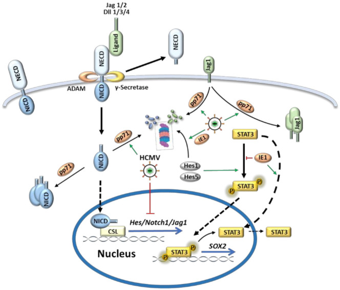
Hearing loss is one of the most prevalent sensory disabilities worldwide with huge social and economic burdens. The leading cause of sensorineural hearing loss (SNHL) in children is congenital cytomegalovirus (CMV) infection. Though the implementation of universal screening and early intervention such as antiviral or anti-inflammatory ameliorate the severity of CMV-associated diseases, direct and targeted therapeutics is still seriously lacking. The major hurdle for it is that the mechanism of CMV induced SNHL has not yet been well understood. In this review, we focus on the impact of CMV infection on the key players in inner ear development including the Wnt and Notch signaling pathways. Investigations on these interactions may gain new insights into viral pathogenesis and reveal novel targets for therapy.
Keywords: Notch signaling pathway; Wnt signaling pathway; congenital cytomegalovirus infection; cytomegalovirus; inner ear development; sensorineural hearing loss.
Conflict of interest statement
The authors declare no conflict of interest.
Figures


Similar articles
-
A wider role for congenital cytomegalovirus infection in sensorineural hearing loss.Pediatr Infect Dis J. 2003 Jan;22(1):39-42. doi: 10.1097/00006454-200301000-00012. Pediatr Infect Dis J. 2003. PMID: 12544407
-
Congenital cytomegalovirus infection: audiologic outcome.Clin Infect Dis. 2013 Dec;57 Suppl 4(Suppl 4):S182-4. doi: 10.1093/cid/cit609. Clin Infect Dis. 2013. PMID: 24257423 Free PMC article.
-
Viral load in children with congenital cytomegalovirus infection identified on newborn hearing screening.J Clin Virol. 2015 Apr;65:41-5. doi: 10.1016/j.jcv.2015.01.015. Epub 2015 Jan 23. J Clin Virol. 2015. PMID: 25766986
-
Congenital cytomegalovirus infection and hearing loss.Herpes. 2005 Oct;12(2):50-5. Herpes. 2005. PMID: 16209862 Review.
-
Evaluation and management of cytomegalovirus-associated congenital hearing loss.Curr Opin Otolaryngol Head Neck Surg. 2017 Oct;25(5):390-395. doi: 10.1097/MOO.0000000000000401. Curr Opin Otolaryngol Head Neck Surg. 2017. PMID: 28857892 Review.
Cited by
-
The Pathogenesis of Cytomegalovirus and Other Viruses Associated with Hearing Loss: Recent Updates.Viruses. 2023 Jun 16;15(6):1385. doi: 10.3390/v15061385. Viruses. 2023. PMID: 37376684 Free PMC article. Review.
-
Congenital CMV infection and central nervous system involvement: mechanisms, treatment, and long-term outcomes.Eur J Pediatr. 2025 May 31;184(6):381. doi: 10.1007/s00431-025-06215-4. Eur J Pediatr. 2025. PMID: 40448827 Review.
-
Development of a Vaccine against Human Cytomegalovirus: Advances, Barriers, and Implications for the Clinical Practice.Vaccines (Basel). 2021 May 25;9(6):551. doi: 10.3390/vaccines9060551. Vaccines (Basel). 2021. PMID: 34070277 Free PMC article. Review.
-
Human Cytomegalovirus IE1 Impairs Neuronal Migration by Downregulating Connexin 43.J Virol. 2023 May 31;97(5):e0031323. doi: 10.1128/jvi.00313-23. Epub 2023 Apr 25. J Virol. 2023. PMID: 37097169 Free PMC article.
References
Publication types
MeSH terms
Substances
LinkOut - more resources
Full Text Sources
Other Literature Sources
Medical
