. 2021 Apr 8;10(4):837.
doi: 10.3390/cells10040837.

Neurodegeneration Induced by Anti-IgLON5 Antibodies Studied in Induced Pluripotent Stem Cell-Derived Human Neurons

Affiliations

Neurodegeneration Induced by Anti-IgLON5 Antibodies Studied in Induced Pluripotent Stem Cell-Derived Human Neurons

Matias Ryding et al. Cells. .

Abstract

Anti-IgLON5 disease is a progressive neurological disorder associated with autoantibodies against a neuronal cell adhesion molecule, IgLON5. In human postmortem brain tissue, the neurodegeneration and accumulation of hyperphosphorylated tau (p-tau) are found. Whether IgLON5 antibodies induce neurodegeneration or neurodegeneration provokes an immune response causing inflammation and antibody formation remains to be elucidated. We investigated the effects of anti-IgLON5 antibodies on human neurons. Human neural stem cells were differentiated for 14-48 days and exposed from Days 9 to 14 (short-term) or Days 13 to 48 (long-term) to either (i) IgG from a patient with confirmed anti-IgLON5 antibodies or (ii) IgG from healthy controls. The electrical activity of neurons was quantified using multielectrode array assays. Cultures were immunostained for β-tubulin III and p-tau and counterstained with 4',6-Diamidine-2'-phenylindole dihydrochloride (DAPI). To study the impact on synapses, cultures were also immunostained for the synaptic proteins postsynaptic density protein 95 (PSD95) and synaptophysin. A lactate dehydrogenase release assay and nuclei morphology analysis were used to assess cell viability. Cultures exposed to anti-IgLON5 antibodies showed reduced neuronal spike rate and synaptic protein content, and the proportion of neurons with degenerative appearance including p-tau (T205)-positive neurons was higher when compared to cultures exposed to control IgG. In addition, cell death was increased in cultures exposed to anti-IgLON5 IgG for 21 days. In conclusion, pathological anti-IgLON5 antibodies induce neurodegenerative changes and cell death in human neurons. This supports the hypothesis that autoantibodies may induce the neurodegenerative changes found in patients with anti-IgLON5-mediated disease. Furthermore, this study highlights the potential use of stem cell-based in vitro models for investigations of antibody-mediated diseases. As anti-IgLON5 disease is heterogeneous, more studies with different IgLON5 antibody samples tested on human neurons are needed.

Keywords: IgLON5; autoimmune encephalitis; neurodegeneration; phosphorylated tau.


Conflict of interest statement

The authors declare no conflict of interest

Figures

Figure 1
Figure 1
Graphical illustration of the experimental setup. Short-term autoantibody exposure was performed on human Neuronal Stem cells (hNSC) and the experimental setup is shown in panel (A). Long-term autoantibody exposure was performed on human-induced Pluripotent Stem Cell (hiPSC)-derived neurons and the experimental setup is shown in panel (B). Black arrows and text specify differentiation procedures. Colored arrows and text boxes specify the time points at which different analyses were performed. LDH: lactate dehydrogenase, MEA: multi electrode array.
Figure 2
Figure 2
Anti-IgLON5 antibodies cause neurodegenerative changes in human neural stem cell derived (hNSC) neural cultures. Anti-IgLON5 antibodies significantly reduced the number of cell surface IgLON5 clusters (A) (n = 6). When evaluating degenerative changes in hNSC-derived cells after 5 days of antibody exposure, there was a tendency toward increased neuronal blebbing (B) (n = 18–19) when comparing anti-IgLON5 IgG to control IgG. There was also a significant difference in fragmented processes (C) (n = 18–19) between anti-IgLON5 IgG and Control IgG-treated groups. Cultures treated with anti-IgLON5 IgG showed an increase in the percentage of neurons with phosphorylated-tau (p-tau) (T205) when compared to cultures exposed to control IgG (D) (n = 18–19). Statistical method: two-tailed unpaired t-tests (* p < 0.05; ** p < 0.01; **** p < 0.0001).
Figure 3
Figure 3
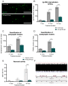
Anti-IgLON5 antibodies reduce cell surface IgLON5 clusters, synaptic proteins, and neuronal activity. Indirect immunofluorescent staining of human induced pluripotent stem cell (hiPSC) cultures treated with control IgG or anti-IgLON5 IgG (A). Quantification of IgLON5-positive clusters in hiPSC cultures revealed a significant reduction after 21 and 35 days of antibody exposure (B) (n = 9). Immunostaining for the synaptic proteins synaptophysin and PSD95 revealed a decrease in synaptophysin clusters after 21 days of exposure and in both synaptophysin and PSD95 clusters after 35 days of exposure (C,D) (n = 6). Multi electrode array (MEA) analysis of hiPSC cultures showed the expected increase in neuronal activity as they matured, however, the spike rate of neurons treated with anti-IgLON5 IgG was significantly lower than that of untreated neurons (E) (n = 5). Example of a MEA data sample is shown in (F) (top). When the signal crosses the threshold (horizontal lines) a spike is registered. Registered spikes from a single electrode in a well with an untreated culture (F, middle) and an anti-IgLON5 IgG-treated culture (F, bottom) during a 15 s period. Statistical analysis: two-tailed unpaired t-test or Two-way ANOVA followed by Sidak’s multiple comparisons test (*: p < 0.05, ***: p < 0.001, ****: p < 0.0001). Scale bar length: 5 μm.
Figure 4
Figure 4
Anti-IgLON5 antibodies increase cell death in induced pluripotent stem cell (hiPSC)-derived neural cultures. Cultures treated with anti-IgLON5 IgG showed an increase in phosphorylated-tau (p-tau) (T205) expression after 7 and 21 days of exposure (AC) (n = 7–8). Quantification of nuclei with unhealthy morphology revealed an increased percentage of dying cells after 21 and 35 days of exposure (D) (n = 7–18). A lactate dehydrogenase (LDH)-assay (a marker of necrosis) showed increased LDH release to the culture medium of anti-IgLON5 IgG exposed cultures after 30 days (E) (n = 8). Statistical analysis: two-way ANOVA followed by Sidak’s multiple comparisons test. (* p < 0.05, ** p < 0.01, *** p < 0.001, **** p < 0.0001). Scale bars: 50 μm. β-tub III: β-tubulin III.

References

    1. Sabater L., Gaig C., Gelpi E., Bataller L., Lewerenz J., Torres-Vega E., Contreras A., Giometto B., Compta Y., Embid C., et al. A novel non-rapid-eye movement and rapid-eye-movement parasomnia with sleep breathing disorder associated with antibodies to IgLON5: A case series, characterisation of the antigen, and post-mortem study. Lancet Neurol. 2014;13:575–586. doi: 10.1016/S1474-4422(14)70051-1. - DOI - PMC - PubMed
    1. Gelpi E., Höftberger R., Graus F., Ling H., Holton J.L., Dawson T., Popovic M., Pretnar-Oblak J., Högl B., Schmutzhard E., et al. Neuropathological criteria of anti-IgLON5-related tauopathy. Acta Neuropathol. 2016;132:531–543. doi: 10.1007/s00401-016-1591-8. - DOI - PMC - PubMed
    1. Gaig C., Graus F., Compta Y., Högl B., Bataller L., Brüggemann N., Giordana C., Heidbreder A., Kotschet K., Lewerenz J., et al. Clinical manifestations of the anti-IgLON5 disease. Neurology. 2017;88:1736–1743. doi: 10.1212/WNL.0000000000003887. - DOI - PMC - PubMed
    1. Nissen M.S., Blaabjerg M. Anti-IgLON5 Disease: A Case With 11-Year Clinical Course and Review of the Literature. Front. Neurol. 2019;10:1056. doi: 10.3389/fneur.2019.01056. - DOI - PMC - PubMed
    1. Cabezudo-García P., Mena-Vázquez N., Torrús G.E., Serrano-Castro P. Response to immunotherapy in anti-IgLON5 disease: A systematic review. Acta Neurol. Scand. 2019;141:263–270. doi: 10.1111/ane.13207. - DOI - PubMed

Publication types

LinkOut - more resources