. 2021 Apr 8;22(8):3851.
doi: 10.3390/ijms22083851.

Differential Therapeutic Effect of Extracellular Vesicles Derived by Bone Marrow and Adipose Mesenchymal Stem Cells on Wound Healing of Diabetic Ulcers and Correlation to Their Cargoes

Affiliations

Differential Therapeutic Effect of Extracellular Vesicles Derived by Bone Marrow and Adipose Mesenchymal Stem Cells on Wound Healing of Diabetic Ulcers and Correlation to Their Cargoes

Margherita Pomatto et al. Int J Mol Sci. .

Abstract

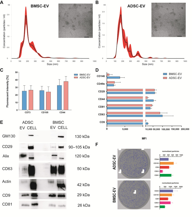
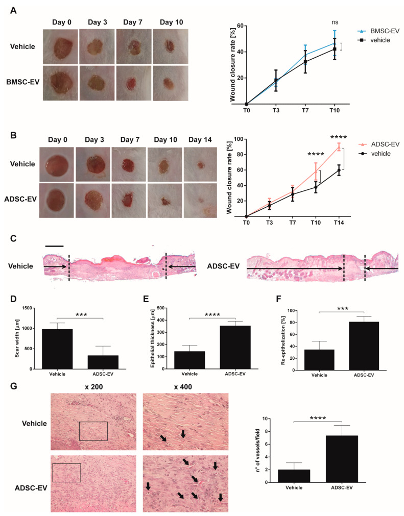
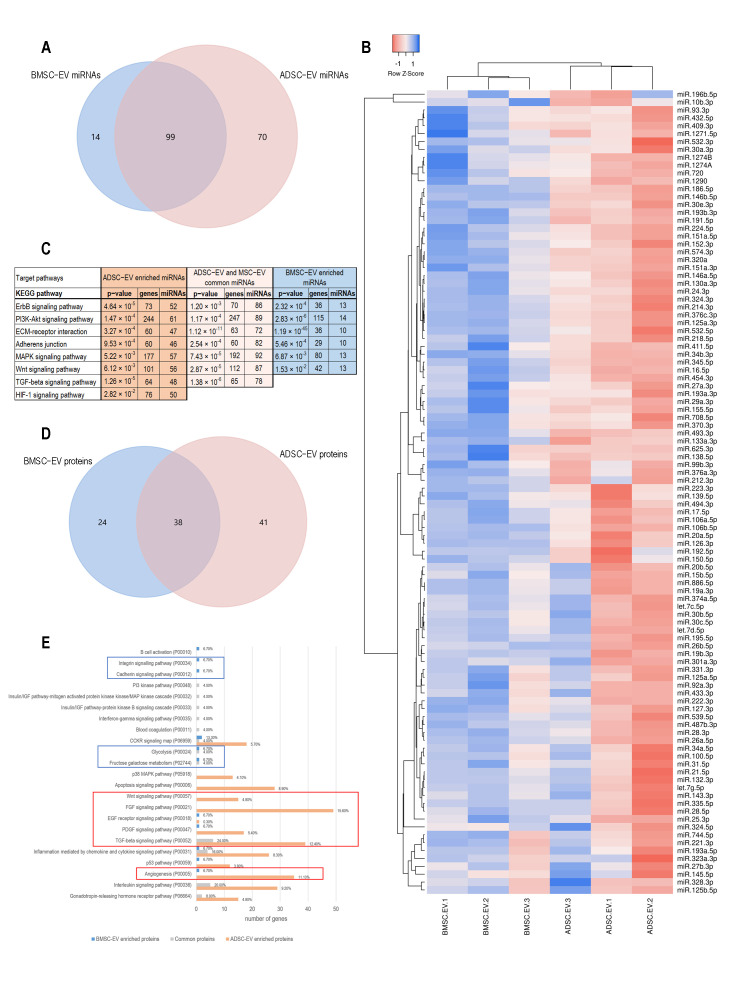
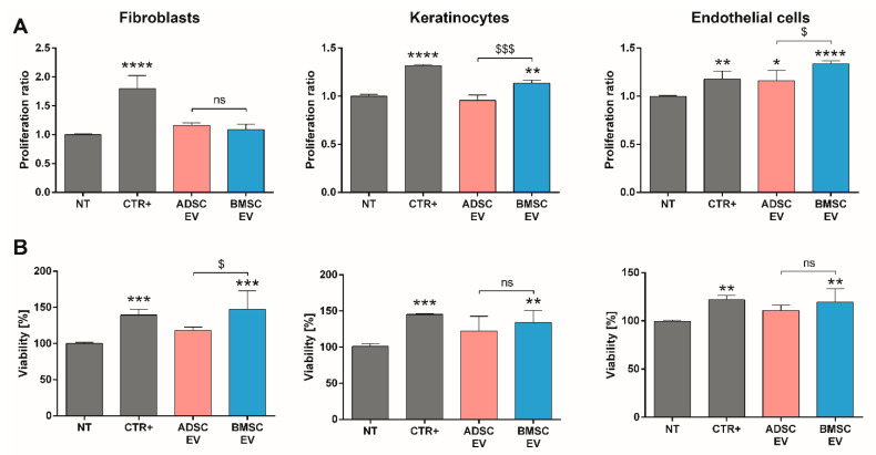
Extracellular vesicles (EVs) derived from mesenchymal stem cells isolated from both bone marrow (BMSCs) and adipose tissue (ADSCs) show potential therapeutic effects. These vesicles often show a similar beneficial effect on tissue regeneration, but in some contexts, they exert different biological properties. To date, a comparison of their molecular cargo that could explain the different biological effect is not available. Here, we demonstrated that ADSC-EVs, and not BMSC-EVs, promote wound healing on a murine model of diabetic wounds. Besides a general similarity, the bioinformatic analysis of their protein and miRNA cargo highlighted important differences between these two types of EVs. Molecules present exclusively in ADSC-EVs were highly correlated to angiogenesis, whereas those expressed in BMSC-EVs were preferentially involved in cellular proliferation. Finally, in vitro analysis confirmed that both ADSC and BMSC-EVs exploited beneficial effect on cells involved in skin wound healing such as fibroblasts, keratinocytes and endothelial cells, but through different cellular processes. Consistent with the bioinformatic analyses, BMSC-EVs were shown to mainly promote proliferation, whereas ADSC-EVs demonstrated a major effect on angiogenesis. Taken together, these results provide deeper comparative information on the cargo of ADSC-EVs and BMSC-EVs and the impact on regenerative processes essential for diabetic wound healing.

Keywords: MSC; adipose; bone marrow; cargo; diabetes; exosome; extracellular vesicle; mesenchymal; therapy; wound healing.


Conflict of interest statement

The authors declare no conflict of interest.

Figures

Figure 1
Figure 1
BMSC and ADSC-EV Characterization. EVs were isolated by BMSCs and ADSCs and analyzed using different techniques. Representative Nanoparticle tracking analyses showing the size distribution and representative Transmission electron microscopy of EVs derived from BMSCs (A) and ADSCs (B) with scale bar 100 nm; (C) flow cytometry analysis (FACS) of EVs for surface proteins CD73, CD105 and CD44 showing the percentage of fluorescent intensity; (D) MACS multiplex bead-based flow cytometry assay of different surface markers, only expressed markers are shown as mean fluorescent intensity (MFI); (E) representative Western blot analysis of integrin β1 (CD29), Actin, exosomal markers CD63, CD9, CD81, Alix, and intracellular marker GM130 as negative control for exosomes in BMSC and ADSC-EVs and cells of origin; (F) Interferometry images of a representative anti-CD9 capture spot post-scan for ADSC-EVs (upper-left) and BMSC-EVs (lower-left). Blue circles indicate EVs detected by interferometry. Histograms show the number of normalized particles counted by interferometry for each capturing antibody (CD9, CD63, CD81, CD44, CD105).
Figure 2
Figure 2
Therapeutic Effect of BMSC and ADSC-EVs on a Mouse Model of Diabetic Ulcers. (A) Representative photographs of full-thickness excisional wound treated with carboxymethylcellulose (vehicle) or carboxymethylcellulose and BMSC-EVs (BMSC-EV) and quantification of wound closure rate at 0, 3, 7 and 10 days expressed as percentage of the original wound size at day 0. ns: not statistically significant difference between treatment with vehicle or BMSC-EV; (B) Representative image of wounds treated with carboxymethylcellulose (vehicle) or carboxymethylcellulose and ADSC-EVs (ADSC-EV) and quantification of wound closure rate at 0, 3, 7, 10 and 14 days expressed as percentage of the original wound size at day 0. ****: p < 0.0001 between treatment with vehicle or ADSC-EV; (C) Transmitted light representative images of H&E-stained sections of wounded skin sections at 14 days treated with carboxymethylcellulose (vehicle) or carboxymethylcellulose and ADSC-EVs (ADSC-EV). The black arrows point out the scar edges. Scale bar 200 μm; Quantitative analysis of the scar width (D), epithelial thickness (E) (µm) and percentage of re-epithelization (F) induced by ADSC-EVs treatment in comparison to treatment with vehicle alone at 14 days. *** p < 0.005, **** p < 0.0001 versus vehicle; (G) Representative images of H&E staining at different magnification and quantification of the number of vessels present in wound sections. Black arrows indicate vessels in the micrograph. ****: p < 0.0001 between treatment with vehicle or ADSC-EV.
Figure 3
Figure 3
Molecular Analysis of ADSC and BMSC-EV Content. (A) The Venn diagram compares the lists of miRNAs carried by ADSC and BMSC-EVs; (B) The heatmap shows the clustering of the groups of miRNAs carried by both ADSC and BMSC-EVs. Row Z score of −1 (red) correlates to low Ct values and Z score of +1 (blue) correlates to high Ct values; (C) The table shows the most relevant results of target pathway analysis for the lists of miRNAs exclusively carried by ADSC-EVs, miRNAs carried by either ADSC and BMSC-EVs, and miRNAs exclusively carried by BMSC-EVs. The first column shows KEGG pathways, the other columns report p-value, the number of target genes in the pathway, and the number of miRNAs; (D) The Venn diagram compares the lists of proteins carried by ADSC and BMSC-EVs; (E) The graph shows the target pathways for the proteins only carried by BMSC-EVs (blue bars), carried by both ADSC and BMSC-EVs (grey bars), and only carried by ADSC-EVs. X axis shows the number of genes involved in each pathway, labels on the right show the percent of gene hits against total number of pathways. Panther pathway code is shown next to the pathway name. Signaling pathways related to ADSC-EV proteins are shown in orange, pathways related to BMSC-EV proteins are shown in blue, pathways related to common proteins are shown in grey. Red and blue boxes highlight pathways mainly associated to ADSC-EVs or BMSC-EVs, respectively.
Figure 4
Figure 4
In Vitro Analysis of MSC-EVs on Proliferation and Viability of Cells Involved in Wound Healing Process. (A) Proliferation assay of different cell types (fibroblasts, keratinocytes, and endothelial cells) after 24 h of treatment detected using BrdU proliferation assay and expressed as ratio with respect to untreated cells (NT); (B) viability assessment on different cell types (fibroblasts, keratinocytes, and endothelial cells) after 48 h of treatment detected using AlamarBlue reagent and expressed as percentage with respect to untreated cells (NT). Cells were treated with following stimuli: DMEM 0% FBS as negative control for untreated cells (NT), DMEM 0% FBS with 50,000 EVs/cell for BMSC or ADSC-EVs (BMSC or ADSC-EV) and different positive controls (CTR+) for each cell type (MCDB131 medium for endothelial cells, FGM-2 Growth Media for fibroblasts and KGMTM Gold Keratinocyte Growth Medium for keratinocytes). Statistical analysis was performed comparing each sample with NT (*: p < 0.05, **: p < 0.01, ***: p < 0.005, ****: p < 0.001) or comparing BMSC-EV and ADSC EV ($: p < 0.05, $$$: p < 0.005, ns: not statistically significant).
Figure 5
Figure 5
In Vitro Analysis of MSC-EVs Activity on Migration of Cells Involved in Wound Healing Process and Vessel Formation. (A) Scratch test assay on different cell types (fibroblasts, keratinocytes and endothelial cells) for measuring the pro-migration effect of MSC-EVs. The wound closure was measured 24 h after treatment and expressed as percentage of the initial wound area; (B) representative micrographs and quantitative analysis of capillary-like structure formation test on endothelial cells treated for 24 h. Capillary-like structures length was measured and expressed as ratio in comparison to untreated cells (NT). Cells were treated with DMEM 0% FBS for untreated cells (NT), DMEM 0% FBS with 50,000 EVs/cell for BMSC or ADSC-EVs (BMSC or ADSC-EV) and different positive controls (CTR+) for each cell type (MCDB131 medium for endothelial cells, FGM-2 Growth Media for fibroblasts, and KGMTM Gold Keratinocyte Growth Medium for keratinocytes). For capillary-like structures formation test, MCDB131 medium was used as CTR1+ and 0.1 µg/mL EGF as CTR2+. Statistical analysis was performed comparing each sample with NT (*: p < 0.05, **: p < 0.01, ***: p < 0.005, ****: p < 0.001) or comparing BMSC-EV and ADSC EV ($: p < 0.05, $$: p < 0.01, ns: not statistically significant).

References

    1. Théry C., Witwer K.W., Aikawa E., Alcaraz M.J., Anderson J.D., Andriantsitohaina R., Antoniou A., Arab T., Archer F., Atkin-Smith G.K., et al. Minimal information for studies of Extracellular vesicles 2018 (MISEV2018): A position statement of the International Society for Extracellular Vesicles and update of the MISEV2014 guidelines. J. Extracell. Vesicles. 2018;7:1535750. doi: 10.1080/20013078.2018.1535750. - DOI - PMC - PubMed
    1. Gould S.J., Raposo G. As we wait: Coping with an imperfect nomenclature for Extracellular Vesicles. J. Extracell. Vesicles. 2013;2 doi: 10.3402/jev.v2i0.20389. - DOI - PMC - PubMed
    1. Colombo M., Raposo G., Théry C. Biogenesis, secretion, and intercellular interactions of exosomes and other Extracellular Vesicles. Annu. Rev. Cell Dev. Biol. 2014;30:255–289. doi: 10.1146/annurev-cellbio-101512-122326. - DOI - PubMed
    1. Bruno S., Kholia S., Deregibus M.C., Camussi G. The Role of Extracellular Vesicles as Paracrine Effectors in Stem Cell-Based Therapies. Adv. Exp. Med. Biol. 2019;1201:175–193. doi: 10.1007/978-3-030-31206-0_9. - DOI - PubMed
    1. Álvarez-Viejo M. Mesenchymal stem cells from different sources and their derived exosomes: A pre-clinical perspective. World J. Stem Cells. 2020;12:100–109. doi: 10.4252/wjsc.v12.i2.100. - DOI - PMC - PubMed