. 2021 Apr 8;10(8):1566.
doi: 10.3390/jcm10081566.

Skin Manifestation of SARS-CoV-2: The Italian Experience

Affiliations

Skin Manifestation of SARS-CoV-2: The Italian Experience

Gerardo Cazzato et al. J Clin Med. .

Abstract

At the end of December 2019, a new coronavirus denominated Severe Acute Respiratory Syndrome Coronavirus 2 (SARS-CoV-2) was identified in Wuhan, Hubei province, China. Less than three months later, the World Health Organization (WHO) declared coronavirus disease-19 (COVID-19) to be a global pandemic. Growing numbers of clinical, histopathological, and molecular findings were subsequently reported, among which a particular interest in skin manifestations during the course of the disease was evinced. Today, about one year after the development of the first major infectious foci in Italy, various large case series of patients with COVID-19-related skin manifestations have focused on skin specimens. However, few are supported by histopathological, immunohistochemical, and polymerase chain reaction (PCR) data on skin specimens. Here, we present nine cases of COVID-positive patients, confirmed by histological, immunophenotypical, and PCR findings, who underwent skin biopsy. A review of the literature in Italian cases with COVID-related skin manifestations is then provided.

Keywords: COVID-19; Italian; SARS-CoV-2; WHO; manifestation; skin.


Conflict of interest statement

The authors declare no conflict of interest.

Figures
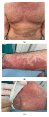
Figure 1
Figure 1
(a) Example of a skin rash with polymorphic erythema characteristics affecting the chest, arms and hands. Erythematous patches with irregular, polycyclic contours. (b) Detail of the erythema on the forearm. (c) Detail of the erythema on the hand. (d) Histopathological findings of erythema-multiforme-like lesions. Note the perivascular lymphocytic infiltrate and focal dermal mucinosis (hematoxylin and eosin (H&E), original magnification 100×). I Details of a SARS-CoV-2 spike protein in endothelial cells from erythema-multiforme-like lesions (immunostaining for SARS-CoV-2 spike proteins, original magnification 400×).
Figure 1
Figure 1
(a) Example of a skin rash with polymorphic erythema characteristics affecting the chest, arms and hands. Erythematous patches with irregular, polycyclic contours. (b) Detail of the erythema on the forearm. (c) Detail of the erythema on the hand. (d) Histopathological findings of erythema-multiforme-like lesions. Note the perivascular lymphocytic infiltrate and focal dermal mucinosis (hematoxylin and eosin (H&E), original magnification 100×). I Details of a SARS-CoV-2 spike protein in endothelial cells from erythema-multiforme-like lesions (immunostaining for SARS-CoV-2 spike proteins, original magnification 400×).
Figure 2
Figure 2
(a) Example of erythematous nodules that were hard, elastic, and painless on palpation, present on the lateral surface of the feet. (b) Histopathological overview of acral lesions (H&E, original magnification, 100×). (c) Details of fragmentation and final dissolution of the collagen and elastic fibers, with the formation of blackish detritus. Presence of reactive fibroblasts. (d) Detail of blood vessels with a prominent endothelium of hobnail type. (H&E, original magnification 400×). (e) Positive immunostaining in eccrine sweat glands (original magnification, 200×).
Figure 2
Figure 2
(a) Example of erythematous nodules that were hard, elastic, and painless on palpation, present on the lateral surface of the feet. (b) Histopathological overview of acral lesions (H&E, original magnification, 100×). (c) Details of fragmentation and final dissolution of the collagen and elastic fibers, with the formation of blackish detritus. Presence of reactive fibroblasts. (d) Detail of blood vessels with a prominent endothelium of hobnail type. (H&E, original magnification 400×). (e) Positive immunostaining in eccrine sweat glands (original magnification, 200×).
Figure 3
Figure 3
Immunohistochemical details of patients with COVID-19-linked maculopapular eruptions (original magnification, 400×).

References

    1. Zhu N., Zhang D., Wang W., Li X., Yang B., Song J., Zhao X., Huang B., Shi W., Lu R., et al. A novel coronavirus from patients with pneumonia in China, 2019. N. Engl. J. Med. 2020;382:727–733. doi: 10.1056/NEJMoa2001017. - DOI - PMC - PubMed
    1. Han Y., Yang H. The transmission and diagnosis of 2019 novel coronavirus infection disease (COVID-19): A Chinese perspective. J. Med. Virol. 2020;92:639–644. doi: 10.1002/jmv.25749. - DOI - PMC - PubMed
    1. World Health Organization Coronavirus Disease (COVID-19)—Events as They Happen. Rolling Updates on Coronavirus Disease (COVID-19) [(accessed on 7 February 2021)];2020 Available online: https://coronavirus.jhu.edu.
    1. Johns Hopkins Corona Resource Center. [(accessed on 7 February 2021)]; Available online: https://coronavirus.jhu.edu.
    1. WHO Coronavirus Diseases. [(accessed on 7 February 2021)]; Available online: https://covid19.who.int/region/euro/country/it.

LinkOut - more resources