Review
. 2021 Apr 8;26(8):2149.
doi: 10.3390/molecules26082149.

Is the Antidepressant Activity of Selective Serotonin Reuptake Inhibitors Mediated by Nicotinic Acetylcholine Receptors?

Affiliations
Review

Is the Antidepressant Activity of Selective Serotonin Reuptake Inhibitors Mediated by Nicotinic Acetylcholine Receptors?

Hugo R Arias et al. Molecules. .

Abstract

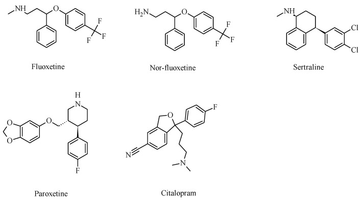
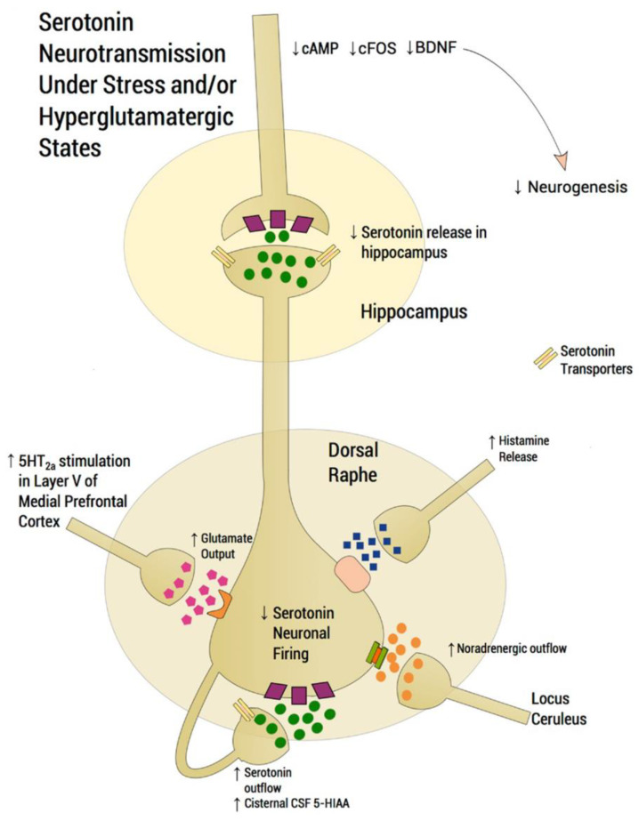
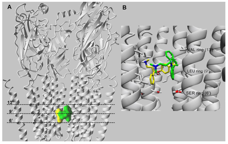
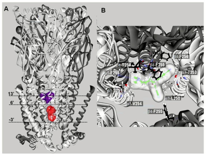
It is generally assumed that selective serotonin reuptake inhibitors (SSRIs) induce antidepressant activity by inhibiting serotonin (5-HT) reuptake transporters, thus elevating synaptic 5-HT levels and, finally, ameliorates depression symptoms. New evidence indicates that SSRIs may also modulate other neurotransmitter systems by inhibiting neuronal nicotinic acetylcholine receptors (nAChRs), which are recognized as important in mood regulation. There is a clear and strong association between major depression and smoking, where depressed patients smoke twice as much as the normal population. However, SSRIs are not efficient for smoking cessation therapy. In patients with major depressive disorder, there is a lower availability of functional nAChRs, although their amount is not altered, which is possibly caused by higher endogenous ACh levels, which consequently induce nAChR desensitization. Other neurotransmitter systems have also emerged as possible targets for SSRIs. Studies on dorsal raphe nucleus serotoninergic neurons support the concept that SSRI-induced nAChR inhibition decreases the glutamatergic hyperstimulation observed in stress conditions, which compensates the excessive 5-HT overflow in these neurons and, consequently, ameliorates depression symptoms. At the molecular level, SSRIs inhibit different nAChR subtypes by noncompetitive mechanisms, including ion channel blockade and induction of receptor desensitization, whereas α9α10 nAChRs, which are peripherally expressed and not directly involved in depression, are inhibited by competitive mechanisms. According to the functional and structural results, SSRIs bind within the nAChR ion channel at high-affinity sites that are spread out between serine and valine rings. In conclusion, SSRI-induced inhibition of a variety of nAChRs expressed in different neurotransmitter systems widens the complexity by which these antidepressants may act clinically.

Keywords: antidepressants; molecular modeling; neuronal pathways; nicotinic acetylcholine receptors; noncompetitive antagonists; selective serotonin reuptake inhibitors.


Conflict of interest statement

The authors declare no conflict of interest. The funders had no role in the design of the study; in the collection, analyses, or interpretation of data; in the writing of the manuscript, or in the decision to publish the results.

Figures

Figure 1
Figure 1
Molecular structures of clinically used selective serotonin reuptake inhibitors (SSRIs) that also inhibit various nicotinic acetylcholine receptor (nAChR) subtypes.
Figure 2
Figure 2
Scheme depicting the functioning of a dorsal raphe nucleus (DRN) serotoninergic neuron under stress conditions that induce depression disorders (modified from [91]). In normal conditions, glutamatergic excitatory afferents from pyramidal neurons of the medial prefrontal cortex (mPFC) directly activate the DRN, thus increasing extracellular serotonin (5-HT), which is regulated by somatodendritic 5-HT1A autoreceptors, decreasing the excessive release in projection areas such as the mPFC and hippocampus. Under stress conditions, however, glutamatergic neurotransmission is hyperactivated, flooding the DRN with 5-HT (i.e., so-called “5-HT flooding”), but concomitantly attenuating 5-HT outflow. An excessive stimulation of 5-HT2ARs in the mPFC increases glutamatergic input, whereas an excessive stimulation of postsynaptic 5-HT1ARs in the hippocampus is associated with a deficit at corticolimbic projection sites, thus compromising neuroplastic processes. Based on this scheme, it is possible that SSRIs alleviate depression symptoms through two processes involving nAChRs: (1) inhibiting presynaptic nAChRs at glutamatergic afferents, which decreases 5-HT flooding to the DRN, and (2) inhibiting postsynaptic nAChRs, which attenuates DRN activity [7,92,93,94]. Since noradrenaline (NE) and histamine are also increased during stress/depression conditions, 5-HT neuron firing is inhibited by NE transmission from the locus coeruleus through α2-heteroceptors and by histamine transmission through histamine-1 receptors. Serotonin transporters are located presynaptically. CSF: cerebrospinal fluid; 5-HIAA: 5-hydroxyindoleacetic acid.
Figure 3
Figure 3
Molecular docking of fluoxetine (in yellow) and imipramine (in green), both in the protonated state, within the α4β2 nAChR ion channel (modified from [70]). (A) Side view of the overlapping binding sites for both ligands that interact with the middle portion of ion channel. (B) Imipramine (in green) and fluoxetine (in yellow) interact with the M2 transmembrane segments forming the lumen of the α4β2 nAChR ion channel. Both ligands interact mainly through van der Waals contacts with a domain formed between the valine (VAL) (position 13′) and serine (SER) (position 6′) rings. In addition, the black arrow indicates the hydrogen bond formed between the oxygen atom of fluoxetine and the hydroxyl group of α4-Ser251 (position 10′). For clarity, one β2 subunit is hidden. Residues involved in ligand binding are presented in stick mode (gray), whereas ligands are rendered either in the ball (A) or stick mode (B). All non-polar hydrogen atoms are hidden.
Figure 4
Figure 4
Docking sites for S-(+)-citalopram (escitalopram) in the (α3)3(β4)2 nAChR model (modified from [68]). (A) Escitalopram docked at two luminal sites (surface model): a high-affinity site located closer to the extracellular ion channel’s mouth (blue) and a low-affinity site located closer to the cytoplasmic side (red). The α3 (white) and β4 (dark gray) subunits are represented as solid ribbons. Dotted lines indicate the positions of the Gly (position -3′), Ser (position 6′), and Val (position 13′) rings along the ion channel. (B) Detailed view of escitalopram at the high-affinity luminal site, showing the cation–π interaction with α3-F255 (position 14′) and the interaction with β4-T254 (position 12′).
Figure 5
Figure 5
Docking sites for S-(+)-citalopram (escitalopram) in the h(α9)2(α10)3 nAChR model (modified from [68]). (A) Escitalopram (light blue surface model) interacted with three possible orthosteric sites located at the interface between the α10(+) (principal component) and α9(−) (or another α10(−)) (complementary component) subunits. The α10 (white) and α9 (dark gray) subunits are represented as solid ribbons. (B) Orthosteric binding sites at the superposed α3(+) (red), α9(+) (black), α10(+) (blue), and α9(−) (white) subunits. Escitalopram is shown as sticks surrounded by the molecular surface. The β9-β10 loop at the α3 and α9 subunits is closer to the receptor center than that at α10, and consequently, there is no room for escitalopram to fit in the agonist binding site in α3 and α9. The α3- and α9-β9-β10 loops overlap the ligand when it is docked as in the α9α10 receptor. (C) Amino acid sequence comparison between α3, α9, and α10 subunits at the level of the β9-β10 loop. Blue: Amino acids identified as causing different β9-β10 loop conformations. (D) Side-chain view showing differences in the occupied volume of side chains at the α10-R186, α9-V186, and α3-Y184 positions, respectively. The side-chain volume differences in the β9-sheet would force the α10-β9-β10 loop to be set apart from the binding pocket.

References

    1. DSM-IV . Diagnostic and Statistical Manual of Mental Disorders IV-TR. American Psychiatric Press; Washington, DC, USA: 2020.
    1. Duman R.S., Aghajanian G.K. Synaptic Dysfunction in Depression: Potential Therapeutic Targets. Science. 2012;338:68–72. doi: 10.1126/science.1222939. - DOI - PMC - PubMed
    1. Salerian A.J., Altar C.A. The Prefrontal Cortex Influence over Subcortical and Limbic Regions Governs Antidepressant Response by N=H/(M+R) Psychiatry Res. 2012;204:1–12. doi: 10.1016/j.pscychresns.2012.04.019. - DOI - PubMed
    1. Zurkovsky L., Taylor W.D., Newhouse P.A. Cognition as a Therapeutic Target in Late-Life Depression: Potential for Nicotinic Therapeutics. Biochem. Pharmacol. 2013;86:1133–1144. doi: 10.1016/j.bcp.2013.07.032. - DOI - PMC - PubMed
    1. Pandya M., Altinay M., Malone D.A., Anand A. Where in the Brain Is Depression? Curr. Psychiatry Rep. 2012;14:634–642. doi: 10.1007/s11920-012-0322-7. - DOI - PMC - PubMed

MeSH terms

Substances

LinkOut - more resources