Case Reports
. 2021 Apr 2;12(4):521.
doi: 10.3390/genes12040521.

MCAT Mutations Cause Nuclear LHON-like Optic Neuropathy

Affiliations
Case Reports

MCAT Mutations Cause Nuclear LHON-like Optic Neuropathy

Sylvie Gerber et al. Genes (Basel). .

Abstract

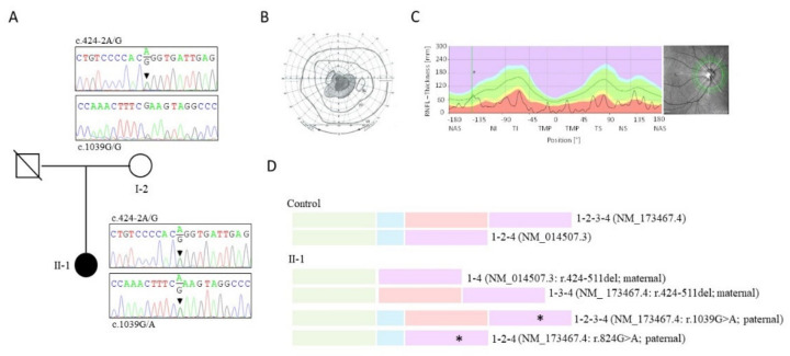
Pathological variants in the nuclear malonyl-CoA-acyl carrier protein transacylase (MCAT) gene, which encodes a mitochondrial protein involved in fatty-acid biogenesis, have been reported in two siblings from China affected by insidious optic nerve degeneration in childhood, leading to blindness in the first decade of life. After analysing 51 families with negative molecular diagnostic tests, from a cohort of 200 families with hereditary optic neuropathy (HON), we identified two novel MCAT mutations in a female patient who presented with acute, sudden, bilateral, yet asymmetric, central visual loss at the age of 20. This presentation is consistent with a Leber hereditary optic neuropathy (LHON)-like phenotype, whose existence and association with NDUFS2 and DNAJC30 has only recently been described. Our findings reveal a wider phenotypic presentation of MCAT mutations, and a greater genetic heterogeneity of nuclear LHON-like phenotypes. Although MCAT pathological variants are very uncommon, this gene should be investigated in HON patients, irrespective of disease presentation.

Keywords: MCAT; hereditary optic neuropathy (HON); nuclear LHON-like.


Conflict of interest statement

The authors declare no conflict of interest.

Figures

Figure 1
Figure 1
Pedigree, ophthalmologic, and genetic data of the Leber hereditary optic neuropathy (LHON)-like malonyl-CoA-acyl carrier protein transacylase (MCAT) case II-1 as follows: (A) Pedigree of the family and electropherograms of exons 2 and 4 showing compound heterozygosity for the NM_173467.4/NM_014507.3:c.424-2A>G and NM_173467.4:c.1039G>A (NM_014507.3:c.824G>A) pathological variants in the affected individual (II-1) and single heterozygosity for the c.424-2A>G mutation in her mother (I-2). The father died before the study. (B) Visual field (right eye) showing central scotoma. (C) Optic coherence tomography of the retinal nerve fibre layer (RNFL) showing marked thinning of the temporal and nasal RNFL. Solid line = II-1; dotted line = controls. Green area = normal RNFL thickness; red area = thickness of abnormally thin RNFL. NAS = nasal; NI = inferior nasal; TI = inferior temporal; TS = superior temporal; NS = superior nasal. (D) Schematic representation of the two MCAT splicing isoforms (with and without exon 3) transcribed from the MCAT gene in the controls, and the four isoforms transcribed in affected individual II-1 from the maternal and paternal alleles, respectively. Each MCAT exon is represented by a colour (green = exon 1; blue = exon 2; red = exon 3; purple = exon 4). The asterisk (*) indicates the paternal mutation in exon 4 (NM_173467.4:c.1039G>A, p.Glu347Lys; NM_014507.3:c.824G>A, p.Glu275Lys). Note that the maternal allele is transcribed into two splicing isoforms lacking exon 23.1.
Figure 2
Figure 2
Structure of human MCAT isoforms and mutant products. The MCAT structure cartoon was prepared with Protein Data Bank (PDB) coordinates for 2C2N (https://www.rcsb.org/) using the PyMOL molecular visualization system, version 1.7.4.0 (Schrödinger, LLC, New York, NY, USA). (A) The full-length protein is composed of a large domain (cyan) and a smaller one (pink). The active-site residues, Ser153, His270, and Arg178, are shown in a gorge between the two subdomains. (B) The protein isoform lacking the amino acid sequence encoded by exon 3 (orange) lacks most of the smaller domain and Arg178, and it is likely inactive. (C) A full and zoom view of Glu347 in the structure. Glu347 is 13.3 Å away from the active-site Ser153. It interacts electrostatically with the side chain of Tyr369 and the backbone amides of Leu318 and Gly319, stabilizing a helix formed by Arg352–Cys361 (green) at the interface between the two domains. (D) The isoform missing residues 142 to 243 (yellow) lacks almost all of the smaller domain, the active-site residues Ser153 and Arg178, and elements of the secondary structure important to the activity of the enzyme.

References

    1. Yu-Wai-Man P., Chinnery P.F. In: Leber Hereditary Optic Neuropathy. Adam M.P., Ardinger H.H., Pagon R.A., Wallace S.E., Bean L.J.H., Mirzaa G., Amemiya A., editors. University of Washington; Seattle, WA, USA: 1993–2021. GeneReviews® [Internet]
    1. Delettre C., Lenaers G., Griffoin J.-M., Gigarel N., Lorenzo C., Belenguer P., Pelloquin L., Grosgeorge J., Turc-Carel C., Perret E., et al. Nuclear gene OPA1, encoding a mitochondrial dynamin-related protein, is mutated in dominant optic atrophy. Nat. Genet. 2000;26:207–210. doi: 10.1038/79936. - DOI - PubMed
    1. Alexander C., Votruba M., Pesch U.E., Thiselton D.L., Mayer S., Moore A., Rodriguez M., Kellner U., Leo-Kottler B., Auburger G., et al. OPA1, encoding a dynamin-related GTPase, is mutated in autosomal dominant optic atrophy linked to chromosome 3q28. Nat. Genet. 2000;26:211–215. doi: 10.1038/79944. - DOI - PubMed
    1. Klebe S., Depienne C., Gerber S., Challe G., Anheim M., Charles P., Fedirko E., Lejeune E., Cottineau J., Brusco A., et al. Spastic paraplegia gene 7 in patients with spasticity and/or optic neuropathy. Brain. 2012;135:2980–2993. doi: 10.1093/brain/aws240. - DOI - PMC - PubMed
    1. Gerber S., Charif M., Chevrollier A., Chaumette T., Angebault C., Kane M.S., Paris A., Alban J., Quiles M., Delettre C., et al. Mutations in DNM1L, as in OPA1, result in dominant optic atrophy despite opposite effects on mitochondrial fusion and fission. Brain. 2017;140:2586–2596. doi: 10.1093/brain/awx219. - DOI - PubMed

Publication types

Substances

LinkOut - more resources