Review
. 2021 Apr 9;11(4):964.
doi: 10.3390/nano11040964.

Silver Nanoparticles as Carriers of Anticancer Drugs for Efficient Target Treatment of Cancer Cells

Affiliations
Review

Silver Nanoparticles as Carriers of Anticancer Drugs for Efficient Target Treatment of Cancer Cells

Helena I O Gomes et al. Nanomaterials (Basel). .

Abstract

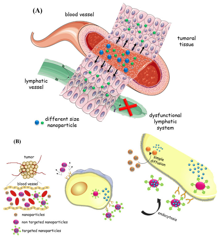
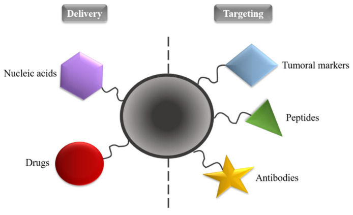
Since the last decade, nanotechnology has evolved rapidly and has been applied in several areas, such as medicine, pharmaceutical, microelectronics, aerospace, food industries, among others. The use of nanoparticles as drug carriers has been explored and presents several advantages, such as controlled and targeted release of loaded or coupled drugs, and the improvement of the drug's bioavailability, in addition to others. However, they also have some limitations, related to their in vivo toxicity, which affects all organs including the healthy ones, and overall improvement in the disease treatment, which can be unnoticeable or minimal. Silver nanoparticles have been increasingly investigated due to their peculiar physical, chemical, and optical properties, which allows them to cover several applications, namely in the transport of drugs to a specific target in the body. Given the limitations of conventional cancer chemotherapy, which include low bioavailability and the consequent use of high doses that cause adverse effects, strategies that overcome these difficulties are extremely important. This review embraces an overview and presentation about silver nanoparticles used as anticancer drug carrier systems and focuses a discussion on the state of the art of silver nanoparticles exploited for transport of anticancer drugs and their influence on antitumor effects.

Keywords: anticancer drugs; antitumor effects; nanocarriers; silver nanoparticles.


Conflict of interest statement

The authors declare no conflict of interest.

Figures

Figure 1
Figure 1
(A) Enhanced permeability and retention effect; (B) Passive (simple diffusion) and active targeting (endocytosis) of nanoparticles.
Figure 2
Figure 2
Applications of noble metal nanoparticles in cancer.
Figure 3
Figure 3
Chemical structures of anticancer drugs (AG) and AgNPs capping agents (HJ) to mediate interaction between AgNPs and anticancer drugs. (A)—methotrexate; (B)—doxorubicin; (C)—alendronate; (D)—epirubicin; (E)—paclitaxel; (F)—imatinib; (G)—gemcitabine; (H)—folic acid; (I)—polyethyleneimine, branched; (J)—graphene oxide.
Figure 4
Figure 4
Release profiles of (a) free MTX, complete after 180 min; and (b) AgNPs-MTX 200, AgNPs-MTX 300, and AgNPs-MTX 400 (200, 300, and 400 refer to different initial MTX/Ag ratios). Reprinted from Ref. [73], with permission from The Royal Society of Chemistry, 2020.
Figure 5
Figure 5
Cell viability studies of cancer cells MCF-7 (A) and HepG2 (B) upon incubation for 48 h with MTX-GO/AgNPs, and other nanocomposites, with and without NIR (808 nm NIR laser, 3.0 W/cm2, 5 min), through MTS assay. Statistical significance analyzed at different levels: * p < 0.05, ** p < 0.01. Reprinted from Ref. [30], with permission from Elsevier, 2021.
Figure 6
Figure 6
Fluorescence microscopy images of cancer cells HepG-2 after incubation with Ag-NGO-DOX (12.5 µg/mL): (a) bright field; (b) blue emission (λex = 360 nm ± 20 nm) of the cell nucleus after staining with 4′,6-diamidino-2-phenylindole (DAPI); (c) red fluorescence emission of DOX inside the cells, by excitation at λex = 490 ± 20 nm; (d) overlapping of (b,c), showing that DOX reached the cancer cells’ nucleus; (e) MTT assay viability results of HepG-2 liver cancer cells after incubation with different concentrations (0, 0.78, 1.56, 3.13, 6.25, 12.5, 25, 50, and 100 µg/mL) of free DOX, Ag-NGO, and Ag-NGO-DOX. Reproduced from [74], with permission from American Chemical Society, 2018.
Figure 7
Figure 7
Influence of pH (37 °C, pH 7.4 (red curve) and pH 5.4 (black curve)) on DOX release profiles from DOX-Ald@AgNPs. Reproduced from Ref. [76] with permission from The Royal Society of Chemistry, 2021.
Figure 8
Figure 8
Influence of AgNPs, Ag@PEI, and Ag@PEI@PTX on the growth of HepG2 cells: (A) at concentrations of 2.5 μg/mL, on the cell viabilities of hepatocarcinoma HepG2 and normal human liver LO2 cell lines, for 24 h by MTT assay; and (B) respective morphological changes. Statistical significance analyzed at different levels: * p < 0.05, ** p < 0.01. Reproduced from Ref. [77], originally from Dove Medical Press Ltd.
Figure 9
Figure 9
Profile of in vitro release of IMAB from IMAB-AgNPs, at 80 h of contact time in phosphate buffer (pH = 7.4), at 37 °C. Reprinted from [40], with permission of the publisher Taylor & Francis Ltd., 2021.
Figure 10
Figure 10
Influence of AgNPs, free IMAB, and IMAB-AgNPs on MCF-7 cancer cells, at different concentrations (0, 1.25, 2.5, 5, 10, 20, and 30 µM) and after incubation of 24 h. Statistical significance analyzed with Student’s t-test at different levels: * p < 0.05, ** p < 0.01 and *** p < 0.001. Reprinted from [40], with permission of the publisher Taylor & Francis Ltd., 2021.
Figure 11
Figure 11
Profiles of GEM release, free in solution and from GEM-AgNPs, in a phosphate buffer solution (pH = 7.4), for 24 h. Reprinted from [59], with permission from Elsevier, 2021.
Figure 12
Figure 12
Cytotoxicity studies with AgNPs (A), free GEM (B), and GEM-AgNPs (C) against the breast cancer cell line MDA-MB-453, at concentrations of 1.56, 3.12, 6.25, 12.5, 25, 50, 75, and 100 µM. (*)—Statistical significance analyzed at the level p < 0.05. Reprinted from [59], with permission from Elsevier, 2021.

References

    1. Azharuddin M., Zhu G.H., Das D., Ozgur E., Uzun L., Turner A.P.F., Patra H.K. A repertoire of biomedical applications of noble metal nanoparticles. Chem. Commun. 2019;55:6964–6996. doi: 10.1039/C9CC01741K. - DOI - PubMed
    1. Zhang X.-F., Liu Z.-G., Shen W., Gurunathan S. Silver Nanoparticles: Synthesis, Characterization, Properties, Applications, and Therapeutic Approaches. Int. J. Mol. Sci. 2016;17:1534. doi: 10.3390/ijms17091534. - DOI - PMC - PubMed
    1. Gali-Muhtasib H., Chouaib R. Nanoparticle Drug Delivery Systems for Cancer Treatment. 1st ed. Jenny Stanford Publishing; Boca Raton, FL, USA: 2020. Nanoparticles in cancer treatment: Types and preparation methods.
    1. Ealia A.M., Saravanakumar M.P. A review on the classification, characterisation, synthesis of nanoparticles and their application. IOP Conf. Ser. Mater. Sci. Eng. 2017;263:032019. doi: 10.1088/1757-899X/263/3/032019. - DOI
    1. Gali-Muhtasib H., Chouaib R. Nanoparticle Drug Delivery Systems for Cancer Treatment. 1st ed. Jenny Stanford Publishing; Boca Raton, FL, USA: 2020. Anti-tumor activity of verbascoside-loaded noble metal nanoparticles.

LinkOut - more resources