. 2021 Apr 9;11(4):1073.
doi: 10.3390/ani11041073.

Prostaglandin F2 Alpha Triggers the Disruption of Cell Adhesion with Cytokeratin and Vimentin in Bovine Luteal Theca Cells

Affiliations

Prostaglandin F2 Alpha Triggers the Disruption of Cell Adhesion with Cytokeratin and Vimentin in Bovine Luteal Theca Cells

Sang-Hee Lee et al. Animals (Basel). .

Abstract

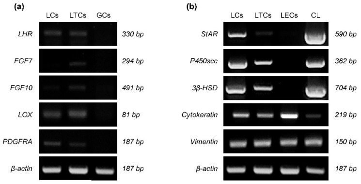
Intermediate filaments (IFs) maintain cell-cell adhesions and are involved in diverse cellular processes such as cytokinesis, cell migration and the maintenance of cell structure. In this study, we investigated the influence of prostaglandin F2 alpha (PGF2α) on cytokeratin and vimentin IFs, Rho-associated protein kinase (ROCK), and cell-cell adhesion in bovine luteal theca cells (LTCs). The luteal cells were isolated from bovine corpus luteum (CL), and the LTCs were treated with 0, 0.01, 0.1 and 1.0 mM PGF2α. Cytokeratin, vimentin and desmoplakin proteins were disrupted and the ROCK protein was significantly increased in PGF2α-treated LTCs. In addition, cell-cell adhesion was significantly (p < 0.05) decreased in the PGF2α-induced LTCs compared to control group (0 mM PGF2α). In conclusion, PGF2α affected the adhesion of cell to cell via disruption of desmoplakin, cytokeratin and vimentin, additionally increasing ROCK in bovine LTCs. These results may provide a better understanding of the mechanism of bovine CL regression.

Keywords: cell adhesion; corpus luteum; luteolysis; ovary; prostaglandin F2 alpha.


Conflict of interest statement

The authors declare no competing interests.

Figures

Figure 1
Figure 1
Isolation of bovine luteal theca cells (LTCs) and detection of morphology using flow cytometry. The corpus luteum (CL) tissues ((a), black cycle) were used at 12–15 days after ovulation and luteal cells ((b); LCs) and LTCs ((f), white area, and (g)) were used for experiment. The LCs and LTCs were stained by β-actin antibody conjugated with conjugated to fluorescein (FITC) dye to distinguish debris from the LCs (c,h). The subpopulation of β-actin-positive LCs ((d), red dots) were largely classified into three areas: red blood cells ((d), yellow area), other luteal cells ((d), green area) and luteal granulosa cells (LGCs; (d), blue area). The relative size of the LCs was analyzed using histogram data of flow cytometry (e). The subpopulation of the β-actin-positive LTCs ((h,i), red dots) was observed. The maximum peaks of x-axis of LTs (e) and LTCs (j) histogram data were matched (Red arrow). White scale bar: 100 μm and yellow scale bar: 400 μm.
Figure 2
Figure 2
Histological images of bovine corpus luteum (CL) section. The CL sections were stained using Eosin-Y and hematoxylin method, secretion-(a) and regression (b)-phases CL were isolated from heifer. White bar: 25 μm.
Figure 3
Figure 3
Expression of mRNA related with theca cells lineage markers (a), steroidogenic function and intermediate filament (b) in bovine luteal theca cells (LTCs). LCa: uteal cells, GCs: granulosa cells, LECs: luteal endothelial cells, CL: secretion-phase corpus luteum (CL), LHR: luteinizing hormone receptor, FGF7: fibroblast growth factor 7, LOX: lysyl oxidase, PDGFRA: platelet derived growth factor receptor alpha, StAR: steroidogenic acute regulatory protein, P450scc: cholesterol side-chain cleavage enzyme, and 3β-HSD: 3β-hydroxysteroid dehydrogenase.
Figure 4
Figure 4
Confocal laser scanning microscope image of cytokeratin protein in prostaglandin F2 alpha (PGF2α)-induced bovine luteal thecal cells (LTCs). The LTCs were incubated in culture media supplemented 0 mM (a,e), 0.01 mM (b,f), 0.1 mM (c,g), and 1.0 mM (d,h) PGF2α. Green: cytokeratin protein, blue: nucleus, yellow arrows: damaged cytokeratin, white line: 50 μm, yellow line: 20 μm.
Figure 5
Figure 5
Confocal laser scanning microscope image of vimentin protein in prostaglandin F2 alpha (PGF2α)-induced bovine luteal thecal cells (LTCs). The LTCs were incubated in culture media supplemented 0 mM (a,e), 0.01 mM (b,f), 0.1 mM (c,g), and 1.0 mM (d,h) PGF2α. Green: vimentin protein, blue: nucleus, white arrows: damaged vimentin, white line: 50 μm, yellow line: 20 μm.
Figure 6
Figure 6
Influence of prostaglandin F2 alpha (PGF2α) on Rho and Rho-associated protein kinase (ROCK) protein in bovine luteal theca cells (LTCs). The protein was analyzed using western blotting. * p < 0.05, n = 5.
Figure 7
Figure 7
Expression of desmoplakin in prostaglandin F2 alpha (PGF2α)-induced bovine luteal thecal cells (LTCs). The LTCs were observed using confocal laser scanning microscope. The LTCs were incubated with 0 mM (a,c) and 0.01 mM (b,d) PGF2α for 24 h, white arrows: normal desmoplakin, yellow arrows: damaged desmoplakin, white line: 50 μm, yellow line: 20 μm.
Figure 8
Figure 8
Effect of prostaglandin F2 alpha (PGF2α) on cell–cell adhesion ability in bovine luteal theca cells (LTCs). The cell-tracker Red stained LTCs were incubated with 0 mM (a) and 0.01 mM (b) PGF2α on the monolayer LTCs. The attached LTCs ((a,b), red dots) on the monolayer cells were counted using the segmentation tools of ImageJ, and the number of attached cells was normalized to 0 M PGF2α treatment (c). * p < 0.05, n = 5.

Similar articles

References

    1. Skarzynski D., Piotrowska-Tomala K., Lukasik K., Galvao A., Farberov S., Zalman Y., Meidan R. Growth and regression in bovine corpora lutea: Regulation by local survival and death pathways. Reprod. Domest. Anim. 2013;48:25–37. doi: 10.1111/rda.12203. - DOI - PubMed
    1. O’shea J., Rodgers R., D’occhio M. Cellular composition of the cyclic corpus luteum of the cow. Reproduction. 1989;85:483–487. doi: 10.1530/jrf.0.0850483. - DOI - PubMed
    1. Groten T., Fraser H., Duncan W., Konrad R., Kreienberg R., Wulff C. Cell junctional proteins in the human corpus luteum: Changes during the normal cycle and after HCG treatment. Hum. Reprod. 2006;21:3096–3102. doi: 10.1093/humrep/del286. - DOI - PubMed
    1. Berisha B., Bridger P., Toth A., Kliem H., Meyer H., Schams D., Pfarrer C. Expression and localization of gap junctional connexins 26 and 43 in bovine periovulatory follicles and in corpus luteum during different functional stages of oestrous cycle and pregnancy. Reprod. Domest. Anim. 2009;44:295–302. doi: 10.1111/j.1439-0531.2008.01068.x. - DOI - PubMed
    1. Sundfeldt K., Piontkewitz Y., Billig H., Hedin L. E-cadherin-catenin complex in the rat ovary: Cell-specific expression during folliculogenesis and luteal formation. J. Reprod. Fertil. 2000;118:375–386. doi: 10.1530/jrf.0.1180375. - DOI - PubMed

LinkOut - more resources