. 2021 Apr 9;11(4):1078.
doi: 10.3390/ani11041078.

Cranial Structure of Varanus komodoensis as Revealed by Computed-Tomographic Imaging

Affiliations

Cranial Structure of Varanus komodoensis as Revealed by Computed-Tomographic Imaging

Sara Pérez et al. Animals (Basel). .

Abstract

This study aimed to describe the anatomic features of the normal head of the Komodo dragon (Varanus komodoensis) identified by computed tomography. CT images were obtained in two dragons using a helical CT scanner. All sections were displayed with a bone and soft tissue windows setting. Head reconstructed, and maximum intensity projection images were obtained to enhance bony structures. After CT imaging, the images were compared with other studies and reptile anatomy textbooks to facilitate the interpretation of the CT images. Anatomic details of the head of the Komodo dragon were identified according to the CT density characteristics of the different organic tissues. This information is intended to be a useful initial anatomic reference in interpreting clinical CT imaging studies of the head and associated structures in live Komodo dragons.

Keywords: Komodo dragon; computed tomography; head.


Conflict of interest statement

The authors declare no conflict of interest.

Figures
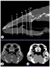
Figure 1
Figure 1
Sagittal image of the head of Varanus komodoensis. The lines and numbers (I–VI) represent the approximate level of the following transverse CT images (C). Transverse CT image of the head at the level of the nasal cavity corresponding to line I. (A) CT bone window. (B) CT soft tissue window. These images are displayed so that the right side of the head is to the viewer’s left and the dorsal view is at the top. N: Nasal bone. Sm: Septomaxilla. Cnd: Dorsal nasal conchae. Cnv: Ventral nasal conchae. Prf: Prefrontal bone. 6. Maxillary bone. T: Tooth. T’: Tooth. Co: Cavum oris. Tg: Tongue. Ms: Musculus intermandibularis + Musculus geniohyoideus + Musculus genioglossus. D: Dentary bone. Ig: Infralabial glands.
Figure 2
Figure 2
Transverse CT image of the head of Varanus komodoensis at the level of the nasal bone corresponding to line II. (A) CT bone window. (B) CT soft tissue window. These images are displayed so that the right side of the head is to the viewer’s left and the dorsal view is at the top. N: Nasal bone. Prf: Prefrontal bone. Ns: Nasal septum. Ng: Nasal glands. St: Stammteil, V: Vomer. Sr: Subconchal recess. Cht: Choanal tube. Nld: Nasolacrimal duct. Mx: Maxillary bone. T: Tooth. Co: Cavum oris. Tg: Tongue. Ms: Musculus intermandibularis + Musculus geniohyoideus + Musculus pterygoideus + Musculus hyoglossus. D: Dentary bone. Mk.fs: Meckelian fossa. Ig: Infralabial glands. Fr: Frontal bone.
Figure 3
Figure 3
Transverse CT image of the head of Varanus komodoensis at the level of the eyes corresponding to line III. (A) CT bone window. (B) CT soft tissue window. These images are displayed so that the right side of the head is to the viewer’s left and the dorsal view is at the top. Frs: Frontal sinus. Br: Brain. Sco: Scleral ossicles. Ls: Lens. Vh: Vitreous humor. Hg: Harderian gland. Sc: Sclera. J: Jugal bone. Ec: Ectopterygoid bone. Pt: Pterygoid bone. D: Dentary bone. Mk.fs: Meckelian fossa. mPt: Musculus pterygoideus. Lxc: Laryngeal cavity. Lxm: Laryngeal muscles. Ms: Musculus intermandibularis + Musculus geniohyoideus + Musculus genioglossus + Musculus hyoglossus. Crc: Cricoid cartilage. Arc: Arytenoid cartilage. Co: Cavum oris. Sr: Sublingual recess.
Figure 4
Figure 4
Transverse CT image of the head of Varanus komodoensis at the level of the parietal bone corresponding to line IV. (A) CT bone window. (B) CT soft tissue window. These images are displayed so that the right side of the head is to the viewer’s left and the dorsal view is at the top. P: Parietal bone (frontoparietal suture). Pfr-Po: Postfrontal + Postorbital bone. Br: Brain. Sp: Sphenoid bone. Pt: Pterygoid bone. San: Surangular bone. Co: Coronoid bone. Co: Cavum oris. Tr: Trachea. Hs: Hyobranchial skeleton. Ms: Musculus intermandibularis + Musculus geniohyoideus + Musculus hyoglossus. J: Jugal bone. Ig: Infralabial glands. mAEM: Musculus adductor mandibulae externus. mPt-mAPM: Musculus pterygoideus + Musculus adductor mandibularis posterior.
Figure 5
Figure 5
Transverse CT image of the head of Varanus komodoensis at the level of the postorbital + postfrontal bone corresponding to line V. (A) CT bone window. (B) CT soft tissue window. These images are displayed so that the right side of the head is to the viewer’s left and the dorsal view is at the top. P: Parietal bone. Lv: Lateral ventricle. Br: Brain. Ep: Epipterygoid bone. Pt: Pterygoid bone. Psp: Parabasisphenoid bone. Pfr-Po: postfrontal + postorbital bone. San: Surangular bone. Co: Cavum oris. Tr: Trachea. Hs: Hyobranchial skeleton. Ms: Musculus intermandibularis + Musculus geniohyoideus + Musculus hyoglossus. mAEM-mPSTs: Musculus adductor mandibulae externus + Musculus pseudotemporalis superficialis. mLPt: Musculus levator pterygoideus. mPt: Musculus pterygoideus.
Figure 6
Figure 6
Transverse CT image of the head of Varanus komodoensis at the level of the squamosal bone corrsponding to line VI. (A) CT bone window. (B) CT soft tissue window. These images are displayed so that the right side of the head is to the viewer’s left and the dorsal view is at the top. Sq: Squamosal bone. P: Parietal bone. Cr: Cerebellum (vermis). Bs: Brain stem. Psp: Parabasisphenoid bone. Pt: Pterygoid bone. San: Surangular bone. Co: Cavum oris. Tr: Trachea. Hs: Hyobranchial skeleton. Ms: Musculus intermandibularis + Musculus geniohyoideus + Musculus hyoglossus. mAEM: Musculus adductor externus mandibularis. mPSTs-p: Musculus pseudotemporalis superficialis and profundus. mPt: Musculus pterygoideus.
Figure 7
Figure 7
Three-dimensional volume-rendered reconstruction image of the cranium of Varanus komodoensis. Dorsal aspect. Pmx: Premaxillary bone. Mx: Maxillary bone. Sm: Septomaxilla. N: Nasal bone. Prf: Prefrontal bone. Fr: Frontal bone. L: Lacrimal bone. J: Jugal bone. Q: Quadrate bone. Sq: Squamosal. Pfr-Po: Postfrontal + postorbital. P: Parietal. Pro: Prootic. San: Surangular bone. Cv1: First cervical vertebra. Cv2: Second cervical vertebra.
Figure 8
Figure 8
Three-dimensional volume-rendered reconstruction image of the cranium of Varanus komodoensis. Ventral aspect. Mx: Maxillary bone. D: Dentary bone. San: Surangular bone. Art: Articular bone. Q: Quadrate bone. Bo. Basioccipital bone. Psp: Parabasisphenoid bone. Pro: Prootic bone. Pt: Pterygoid bone. Pa: Palatine bone. V: Vomer. Sm: Septomaxilla. N: Nasal bone. Fr: Frontal bone. P: Parietal bone. Ha: Hyoid apparatus. Cv2: Second cervical vertebra.
Figure 9
Figure 9
Three-dimensional volume-rendered reconstruction image of the cranium of Varanus komodoensis. Lateral aspect. Pmx: Premaxillary bone. Mx: Maxillary bone. Prf: Prefrontal bone. N: Nasal bone. Fr: Frontal bone. L: Lacrimal bone. J: Jugal bone. Ec: Ectopterygoid bone. Pt: Pterygoid bone. Ep: Epipterygoid. Q: Quadrate bone. Sq: Squamosal. Pfr-Po: Postfrontal + postorbital. Pro: Prootic. D: Dentary bone. T: Tooth. San: Surangular bone. Art: Articular bone. Co: Coronoid bone. Ha: Hyoid apparatus.
Figure 10
Figure 10
Dorsal MIP image of the cranium of Varanus komodoensis. Px: Premaxillary bone. Mx: Maxillary bone. Sm: Septomaxilla. V: Vomer. N: Nasal bone. Prf: Prefrontal bone. Fr: Frontal bone. L: Lacrimal bone. Sco: Scleral ossicles. J: Jugal bone. Pfr-Po: Postfrontal + postorbital. P: Parietal. So: Supraoccipital. Sq: Squamosal. Ost: Osteoderms. Cv1: First cervical vertebra. Cv2: Second cervical vertebra.
Figure 11
Figure 11
Ventral MIP image of the cranium of Varanus komodoensis. D: Dentary bone. Mx: Maxillary bone. San: Surangular bone. Art: Articular bone. Bo. Basioccipital bone. Psp: Parabasisphenoid bone. Oc: Occipital condyle. Pro: Prootic bone. Sm: Septomaxilla. V: Vomer. Pa: Palatine bone. Pt: Pterygoid bone. Cv1: First cervical vertebra. Cv2: Second cervical vertebra.

Similar articles

Cited by

References

    1. Banzato T., Russo E., Di Toma A., Palmisano G., Zotti A. Anatomic imaging of the Boa constrictor head: A comparison between radiography, computed tomography and cadaver anatomy. Am. J. Vet. Res. 2011;72:1592–1599. doi: 10.2460/ajvr.72.12.1592. - DOI - PubMed
    1. Lauridsen H., Hansen K., Wang T., Agger P., Andersen J.L., Knudsen P.S., Rasmussen A.S., Uhrenholt L., Pedersen M. Inside Out: Modern Imaging Techniques to Reveal Animal Anatomy. PLoS ONE. 2011;6:e17879. doi: 10.1371/journal.pone.0017879. - DOI - PMC - PubMed
    1. Deem S.L. Role of the zoo veterinarian in the conservation of captive and free ranging wildlife. Int. Zoo Yearb. 2007;41:3–11. doi: 10.1111/j.1748-1090.2007.00020.x. - DOI
    1. IUCN/SSC IUCN Red List of Threatened Species. [(accessed on 1 December 2020)];2002 Available online: www.redlist.org.
    1. Banzato T., Selleri P., Veladiano I.A., Martin A., Zanetti E., Zotti A. Comparative evaluation of the cadaveric, radiographic and computed tomographic anatomy of the heads of green iguana (Iguana iguana), common tegu (Tupinambis merianae) and bearded dragon (Pogona vitticeps) BMC Vet. Res. 2012;11:53. doi: 10.1186/1746-6148-8-53. - DOI - PMC - PubMed

LinkOut - more resources