. 2021 Apr 18;11(4):1161.
doi: 10.3390/ani11041161.

Remarkable Cryptic Diversity of Paratylenchus spp. (Nematoda: Tylenchulidae) in Spain

Affiliations

Remarkable Cryptic Diversity of Paratylenchus spp. (Nematoda: Tylenchulidae) in Spain

Ilenia Clavero-Camacho et al. Animals (Basel). .

Abstract

In previous studies, fifteen species of Paratylenchus, commonly known as pin nematodes, have been reported in Spain. These plant-parasitic nematodes are ectoparasites with a wide host range and global distribution. In this research, 27 populations from twelve Paratylenchus species from 18 municipalities in Spain were studied using morphological, morphometrical and molecular data. This integrative taxonomic approach allowed the identification of twelve species, four of them were considered new undescribed species and eight were already known described. The new species described here are P. caravaquenus sp. nov., P. indalus sp. nov., P. pedrami sp. nov. and P. zurgenerus sp. nov. As for the already known described species, five were considered as first reports for the country, specifically P.enigmaticus, P. hamatus, P. holdemani, P. israelensis, and P. veruculatus, while P. baldaccii, P. goodeyi and P. tenuicaudatus had already been recorded in Spain. This study provides detail morphological and molecular data, including the D2-D3 expansion segments of 28S rRNA, ITS rRNA, and partial mitochondrial COI regions for the identification of different Paratylenchus species found in Spain. These results confirm the extraordinary cryptic diversity in Spain and with examples of morphostatic speciation within the genus Paratylenchus.

Keywords: D2-D3 of 28S rRNA; ITS rRNA; cytochrome c oxidase subunit 1; molecular; morphology; phylogeny; rRNA; taxonomy.


Conflict of interest statement

The authors declare no conflict of interest. The funders had no role in the design of the study; in the collection, analyses, or interpretation of data; in the writing of the manuscript, or in the decision to publish the results.

Figures
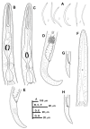
Figure 1
Figure 1
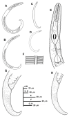
Line drawings of Paratylenchus caravaquenus sp. nov. (A): Entire females and males; (B,C): Female pharyngeal region; (D,E): Female posterior region; (F): Male pharyngeal region showing absence of stylet; (G,H): Male posterior region.
Figure 2
Figure 2
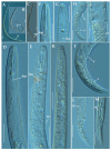
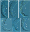
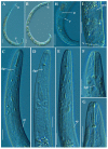
Light photomicrographs of Paratylenchus caravaquenus sp. nov. (A,B): Entire female with vulva arrowed; (C): Entire male with spicules arrowed; (D,E): Female pharyngeal region; (F,G): Female lip region; (H,I): Female posterior region with vulva and anus (arrowed); (J): Detail of vulva showing advulval flap (arrowed); (K): Male pharyngeal region showing absence of stylet; (L,M): Male posterior region showing spicules (arrowed). Scale bars (AI, KM = 20 μm; J = 10 μm). (Abbreviations: a = anus; avf = advulval flap; dgo = pharyngeal dorsal gland orifice; ep = excretory pore; sp = spicules; V = vulva).
Figure 3
Figure 3
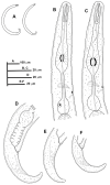
Line drawings of Paratylenchus indalus sp. nov. (AD): Entire females; (E): Female pharyngeal region; (F): Lateral field at mid-body; (G,H): Female posterior region.
Figure 4
Figure 4
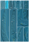
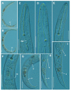
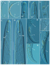
Light photomicrographs of female of Paratylenchus indalus sp. nov. (A,B): Entire female with vulva arrowed; (C,D): Female pharyngeal region; (EH): Female lip region; (I): Fourth-stage juvenile showing stylet (arrowed); (J): Female posterior region with vulva and anus (arrowed); (K): Detail of vulva showing advulval flap (arrowed); (L,M): Female posterior region with lateral field, vulva and anus (arrowed). Scale bars (AD, JM = 20 μm; EI = 10 μm). (Abbreviations: a = anus; avf= advulval flap; dgo = pharyngeal dorsal gland orifice; ep = excretory pore; lf = lateral field; V = vulva).
Figure 5
Figure 5
Light photomicrographs of Paratylenchus indalus sp. nov. female posterior regions (AF). Scale bar: 20 μm). (Abbreviations: a = anus; V = vulva).
Figure 6
Figure 6
Line drawings of Paratylenchus pedrami sp. nov. (A): Entire females; (B,C): Female pharyngeal region; (DH): Female posterior region; (I): Male pharyngeal region showing absence of stylet; (J): Male posterior region.
Figure 7
Figure 7
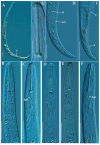
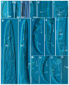
Light photomicrographs of female of Paratylenchus pedrami sp. nov. (A): Entire female with vulva arrowed; (B,C): Female pharyngeal region; (D): Male pharyngeal region; (E,F): Female lip region; (G): Vulval region showing spermatheca (arrowed); (H): Female posterior region showing complete genital branch; (I): Detail of lateral field at mid-body (arrowed); (J): Female posterior region with vulva and anus arrowed; K, L: Male posterior region with spicules (arrowed). Scale bars (A,H,J = 20 μm; BG,I,K,L = 10 μm). (Abbreviations: a = anus; avf = advulval flap; dgo = pharyngeal dorsal gland orifice; ep = excretory pore; lf = lateral field; ov = ovary; spm = spermatheca; V = vulva).
Figure 8
Figure 8
Light photomicrographs of Paratylenchus pedrami sp. nov. female posterior regions (AF). Scale bars (AF = 20 μm). (Abbreviations: a = anus; V = vulva).
Figure 9
Figure 9
Line drawings of Paratylenchus zurgenerus sp. nov. (A): Entire females; (B,C): Female pharyngeal region; (DF): Female posterior region.
Figure 10
Figure 10
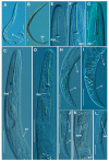
Light photomicrographs of female of Paratylenchus zurgenerus sp. nov. (A,B): Entire female with vulva arrowed; (CE): Female pharyngeal region; (FK): Female lip region; (L): Female posterior region showing empty spermatheca, vulva and anus (arrowed); (M): Detail of vulva showing advulval flap (arrowed); (N): Female mid-region with lateral field (arrowed). Scale bars (AE,L = 20 μm; FK,M,N = 10 μm). (Abbreviations: a = anus; avf = advulval flap; dgo = pharyngeal dorsal gland orifice; ep = excretory pore; lf = lateral field; spm = spermatheca; V = vulva).
Figure 11
Figure 11
Light photomicrographs of Paratylenchus zurgenerus sp. nov. female posterior regions (AF). Scale bars (AF = 20 μm). (Abbreviations: a = anus; V = vulva).
Figure 12
Figure 12
Light photomicrographs of Paratylenchus enigmaticus Munawar, Yevtushenko, Palomares-Rius & Castillo, 2021. (A): Entire female with vulva arrowed; (B,C): Female pharyngeal region; (D,E): Female posterior region showing vulva and anus (arrowed). Scale bars (AE = 20 μm). (Abbreviations: a = anus; avf = advulval flap; dgo = pharyngeal dorsal gland orifice; V= vulva).
Figure 13
Figure 13
Light photomicrographs of Paratylenchus goodeyi (Oostenbrink, 1953) Raski, 1962. (A,B): Entire female with vulva arrowed; (CE): Female lip region; (F): Detail of excretory pore; (GI): Female posterior region with vulva and anus (arrowed). Scale bars (AI = 20 μm). (Abbreviations: a = anus; dgo = pharyngeal dorsal gland orifice; ep = excretory pore; V = vulva).
Figure 14
Figure 14
Light photomicrographs of Paratylenchus baldaccii Raski, 1975. (A): Entire female with vulva arrowed; (BF): Female pharyngeal region; (G): Detail of female stylet; (H): Female posterior region showing lateral field and advulval flap (arrowed); (I): Female posterior region showing vulva and anus (arrowed). Scale bars (AF, H,I = 20 μm; G = 10 μm). (Abbreviations: a = anus; avf = advulval flap; dgo = pharyngeal dorsal gland orifice; lf = lateral field; V = vulva).
Figure 15
Figure 15
Light photomicrographs of Paratylenchus hamatus Thorne & Allen, 1950. (A): Entire female with vulva arrowed; (B): Entire male with spicules arrowed; (C,D): Female pharyngeal region; (EG): Female lip region; (HJ): Female posterior region with vulva and anus (arrowed); (K): Detail of vulva showing spermatheca and advulval flap (arrowed); (L): Male tail with spicules arrowed. Scale bars (AL = 20 μm). (Abbreviations: a = anus; avf = advulval flap; dgo = pharyngeal dorsal gland orifice; ep = excretory pore; spm = spermatheca; sp = spicules; V = vulva).
Figure 16
Figure 16
Light photomicrographs of Paratylenchus holdemani Raski, 1975. (A,B): Entire female with vulva arrowed; (C): Entire male with spicules arrowed; (D): Female pharyngeal region; E: Female lip region; F: Female posterior region with vulva and anus (arrowed); (G): Detail of vulva showing advulval flap (arrowed); (H): Detail of female tail tip; (I): Female posterior region showing lateral field and advulval flap (arrowed); (J): Male tail with spicules arrowed. Scale bars (AF,I,J = 20 μm; GH = 10 μm). (Abbreviations: a= anus; avf= advulval flap; dgo= pharyngeal dorsal gland orifice; ep = excretory pore; lf = lateral field; sp = spicules; V = vulva).
Figure 17
Figure 17
Light photomicrographs of Paratylenchus israelensis (Raski, 1973) Siddiqi, 1986. (A,B): Entire female with vulva arrowed; (C,D): Female pharyngeal region; (E,F): Female lip region; (G): Detail of lateral field at mid-body; (H,J): Female posterior region with vulva and anus (arrowed); (I): Detail of vulva showing advulval flap (arrowed); (K): Detail of female tail tip with anus arrowed. Scale bars (AK = 20 μm). (Abbreviations: a = anus; avf = advulval flap; dgo = pharyngeal dorsal gland orifice; lf = lateral field; V = vulva).
Figure 18
Figure 18
Light photomicrographs of Paratylenchus tenuicaudatus Wu, 1961. (A): Entire female with vulva arrowed; (B,C): Female pharyngeal region; (DG): Female lip region; (H): Detail of female lateral field at mid-body (arrowed); (I): Detail of vulva showing a frontal view of advulval flap (arrowed); (JO): Female posterior region showing vulva and anus (arrowed); (P): Male tail with spicules arrowed. Scale bars (AP = 20 μm). (Abbreviations: a = anus; avf = advulval flap; dgo = pharyngeal dorsal gland orifice; ep = excretory pore; lf = lateral field; spm = spermatheca; sp= spicules; V = vulva).
Figure 19
Figure 19
Light photomicrographs of female of Paratylenchus veruculatus Wu, 1962. (A): Entire female with stylet and vulva arrowed; (B): Fourth-stage juvenile with stylet arrowed; (CE): Female pharyngeal region; (FG): Female lip region; (H): Female posterior region with lateral field and vulva arrowed; (I): Detail of empty spermatheca (arrowed). Scale bars (A,B,H = 20 μm; CG,I= 10 μm). (Abbreviations: a = anus; dgo = pharyngeal dorsal gland orifice; ep = excretory pore; lf = lateral field; spm = spermatheca; st = stylet; V = vulva).
Figure 20
Figure 20
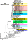
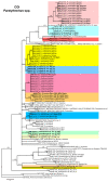
Phylogenetic relationships within the genus Paratylenchus. Bayesian 50% majority rule consensus tree as inferred from D2-D3 expansion domains of the 28S rRNA sequence alignment under the general time-reversible model of sequence evolution with correction for invariable sites and a gamma-shaped distribution (GTR + I + G). Posterior probabilities of more than 0.70 are given for appropriate clades. Newly obtained sequences in this study are shown in bold. The scale bar indicates expected changes per site.
Figure 21
Figure 21
Phylogenetic relationships within the genus Paratylenchus. Bayesian 50% majority rule consensus tree as inferred from ITS rRNA sequence alignment under the general time-reversible model of sequence evolution with correction for invariable sites and a gamma-shaped distribution (GTR + I + G). Posterior probabilities of more than 0.70 are given for appropriate clades. Newly obtained sequences in this study are shown in bold. The scale bar indicates expected changes per site.
Figure 22
Figure 22
Phylogenetic relationships within the genus Paratylenchus. Bayesian 50% majority rule consensus tree as inferred from COI sequence alignment under the general time-reversible model of sequence evolution with a gamma-shaped distribution (GTR + G). Posterior probabilities of more than 0.70 are given for appropriate clades. Newly obtained sequences in this study are shown in bold. The scale bar indicates expected changes per site.

References

    1. Micoletzky H. Die Freilebenden Erd-Nematoden. Archiv für Naturgeschichte; Berlin, Germany: 1922.
    1. Ghaderi R., Geraert E., Karegar A. The Tylenchulidae of the World, Identification of the Family Tylenchulidae (Nematoda: Tylenchida) Academia Press; Ghent, Belgium: 2016.
    1. Ghaderi R. The damage potential of pin nematodes, Paratylenchus Micoletzky, 1922 sensu lato spp. (Nematoda: Tylenchulidae) J. Crop Prot. 2019;8:243–257.
    1. Bongers T., Ferris H. Nematode community structure as a bioindicator in environmental monitoring. Trends Ecol. Evol. 1999;14:224–228. doi: 10.1016/S0169-5347(98)01583-3. - DOI - PubMed
    1. Schmidt J.H., Seeger J.N., Von Grafenstein K., Wintzer J., Finckh M.R., Hallmann J. Population dynamics and host range of Paratylenchus bukowinensis. Nematology. 2020;22:257–267. doi: 10.1163/15685411-00003304. - DOI

LinkOut - more resources