. 2021 Apr 18;9(4):437.
doi: 10.3390/biomedicines9040437.

Bromodomain and Extraterminal Protein Inhibitor, Apabetalone (RVX-208), Reduces ACE2 Expression and Attenuates SARS-Cov-2 Infection In Vitro

Affiliations

Bromodomain and Extraterminal Protein Inhibitor, Apabetalone (RVX-208), Reduces ACE2 Expression and Attenuates SARS-Cov-2 Infection In Vitro

Dean Gilham et al. Biomedicines. .

Abstract

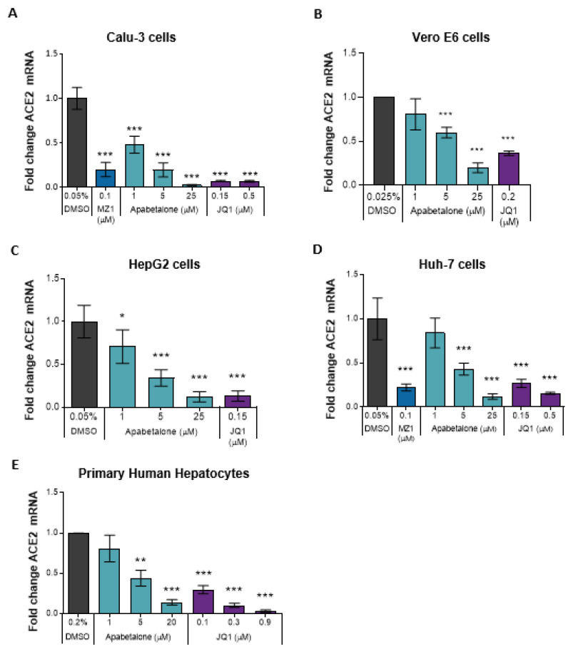
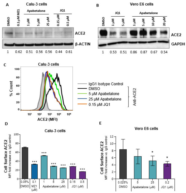
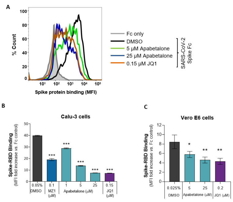
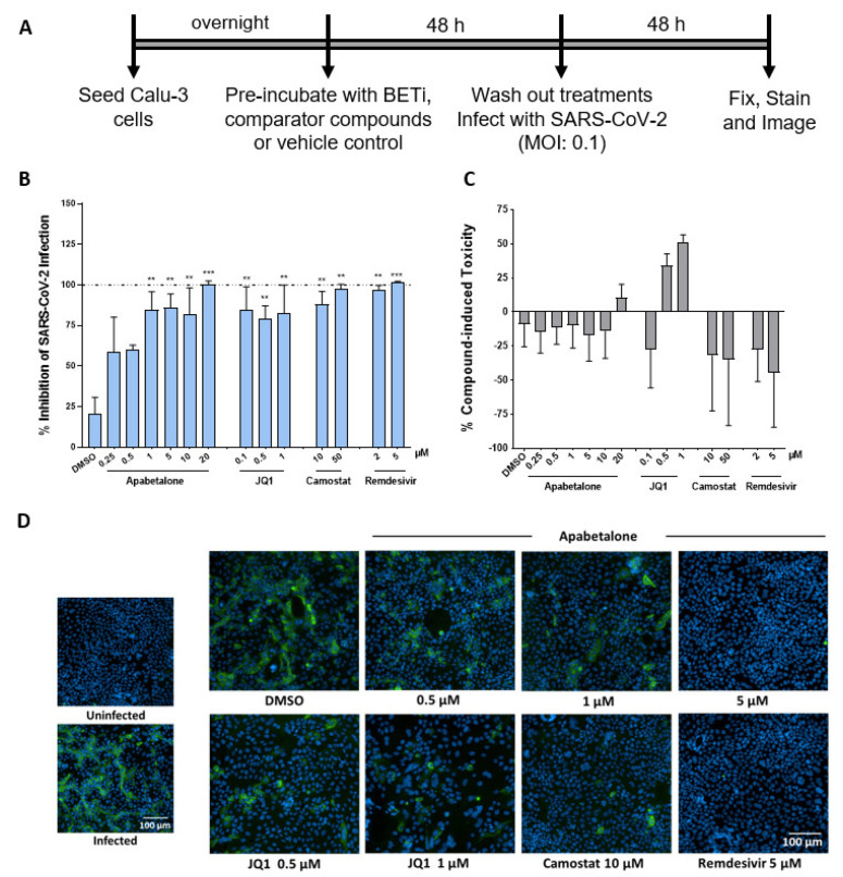
Effective therapeutics are urgently needed to counter infection and improve outcomes for patients suffering from COVID-19 and to combat this pandemic. Manipulation of epigenetic machinery to influence viral infectivity of host cells is a relatively unexplored area. The bromodomain and extraterminal (BET) family of epigenetic readers have been reported to modulate SARS-CoV-2 infection. Herein, we demonstrate apabetalone, the most clinical advanced BET inhibitor, downregulates expression of cell surface receptors involved in SARS-CoV-2 entry, including angiotensin-converting enzyme 2 (ACE2) and dipeptidyl-peptidase 4 (DPP4 or CD26) in SARS-CoV-2 permissive cells. Moreover, we show that apabetalone inhibits SARS-CoV-2 infection in vitro to levels comparable to those of antiviral agents. Taken together, our study supports further evaluation of apabetalone to treat COVID-19, either alone or in combination with emerging therapeutics.

Keywords: BET proteins; COVID-19; SARS-CoV-2; angiotensin-converting enzyme 2 (ACE2); apabetalone.


Conflict of interest statement

A.L.S., D.Y.M., A.M., S.P.M.R., and D.E.-G. have no conflicts of interest, financial or otherwise. D.G., L.F., S.C.S., J.O.J., M.S., N.C.W.W. and E.K. are employed by Resverlogix and may hold stock and/or stock options.

Figures

Figure 1
Figure 1
BETi treatment downregulated ACE2 transcript levels in various cell types. Gene expression of hACE2 (A,CE) or RhACE2 (B) was measured by TaqMan real-time PCR. Calu-3 (A) were treated for 48 h, Vero E6 (B) were treated for 24 h, HepG2 (C), and Huh-7 (D) cells were treated for 96 h. Primary human hepatocytes (n = 3 donors) (E) were treated for 48 h. Error bars represent SD. * p < 0.05, ** p < 0.01, *** p < 0.001, one-way ANOVA followed by Dunnett’s multiple comparison test.
Figure 2
Figure 2
BETi treatment reduced both surface and total ACE2 protein levels. (A) Immunoblot of total ACE2 protein in Calu-3 cells following BETi treatment for 48 h. Values below the blots indicate band quantification via densitometric analysis (ACE2/β-ACTIN) and are represented as fold change to vehicle-treated cells (0.05% v/v DMSO). (B) Immunoblot of total ACE2 protein in Vero E6 cells following BETi treatment for 48 h. Values below the blot indicate band quantification via densitometric analysis (ACE2/GAPDH) and are represented as fold change to vehicle-treated cells (0.1% v/v DMSO). (C) Representative histogram from flow cytometry showing overlay of cell surface ACE2 on Calu-3 cells following 48 h of the indicated treatments. (DE) Quantification of ACE2 protein levels on Calu-3 (D; n = 3) or Vero E6 (E, n = 5) following BETi treatment for 48 h. ACE2 surface expression is shown as mean fluorescent intensity (MFI) ratio to isotype control (DE). Error bars represent SD. * p < 0.05, *** p < 0.001, one-way ANOVA followed by Dunnett’s multiple comparison test.
Figure 3
Figure 3
BET inhibition decreased SARS-CoV-2 spike RBD binding to Calu-3 and Vero E6. (A) A histogram overlay representing the binding of SARS-CoV-2 spike RBD Fc fusion protein to Calu-3 following 48 h of apabetalone or JQ1 treatment at the indicated concentrations. (B,C) Quantification of BETi-driven changes to SARS-CoV-2 spike RBD binding to Calu-3 (B; n = 3) or Vero E6 (C; n = 3) cells following 48 h of treatment. Binding results are shown as mean fluorescent intensity (MFI) ratio to Fc control. Error bars represent SD. * p < 0.05, ** p < 0.01, *** p < 0.001, one-way ANOVA followed by Dunnett’s multiple comparison test.
Figure 4
Figure 4
BETi compounds diminished SARS-CoV-2 infection in Calu-3. (A) Schematic illustration of SARS-CoV-2 infection assay design. Briefly, cells were pre-treated with BETi (apabetalone or JQ1), comparator/control compounds (camostat mesylate (camostat or remdesivir) or vehicle control (DMSO 0.1% v/v) for 48 h. Treatments were washed off and cells were then infected with SARS-CoV-2 at an MOI of 0.1. 48 h post-infection, Calu-3 cells were fixed and stained for spike protein. (B) Percent inhibition of viral infectivity by BETi pre-treatment in Calu-3 cells (blue bars). (C) Compound-induced cytotoxicity in viral infected Calu-3 cells is shown in grey bars (n = 3 independent experiments). (D) Representative immunofluorescence images of virus infection in Calu-3 cells for the treatments and indicated concentrations. Scale bar = 100 μm. Error bars represent SEM. ** p < 0.01, *** p < 0.001, one-way ANOVA followed by Dunnett’s multiple comparison test.

References

    1. Soy M., Keser G., Atagündüz P., Tabak F., Atagündüz I., Kayhan S. Cytokine storm in COVID-19: Pathogenesis and overview of anti-inflammatory agents used in treatment. Clin. Rheumatol. 2020;39:2085–2094. doi: 10.1007/s10067-020-05190-5. - DOI - PMC - PubMed
    1. The World Health Organization: Statement on the second meeting of the International Health Regulations (2005) Emergency Committee regarding the outbreak of novel coronavirus (2019-nCoV) [(accessed on 17 April 2021)]; Available online: https://www.who.int/news/item/30-01-2020-statement-on-the-second-meeting...
    1. Stawicki S.P., Jeanmonod R., Miller A.C., Paladino L., Gaieski D.F., Yaffee A.Q., De Wulf A., Grover J., Papadimos T.J., Bloem C., et al. The 2019–2020 novel coronavirus (severe acute respiratory syndrome coronavirus 2) pandemic: A joint american college of academic international medicine-world academic council of emergency medicine multidisciplinary COVID-19 working group consensus paper. J. Glob. Infect. Dis. 2020;12:47–93. doi: 10.4103/jgid.jgid_86_20. - DOI - PMC - PubMed
    1. Dong Y., Mo X., Hu Y., Qi X., Jiang F., Jiang Z., Tong S. Epidemiology of COVID-19 among children in China. Pediatrics. 2020;145:e20200702. doi: 10.1542/peds.2020-0702. - DOI - PubMed
    1. Barker-Davies R.M., O’Sullivan O., Senaratne K.P.P., Baker P., Cranley M., Dharm-Datta S., Ellis H., Goodall D., Gough M., Lewis S., et al. The Stanford Hall consensus statement for post-COVID-19 rehabilitation. Br. J. Sports Med. 2020;54:949–959. doi: 10.1136/bjsports-2020-102596. - DOI - PMC - PubMed

LinkOut - more resources