. 2021 Apr 18;13(8):1945.
doi: 10.3390/cancers13081945.

The New Serum-Free OptiPASS® Medium in Cold and Oxygen-Free Conditions: An Innovative Conservation Method for the Preservation of MDA-MB-231 Triple Negative Breast Cancer Spheroids

Affiliations

The New Serum-Free OptiPASS® Medium in Cold and Oxygen-Free Conditions: An Innovative Conservation Method for the Preservation of MDA-MB-231 Triple Negative Breast Cancer Spheroids

Antoine Goisnard et al. Cancers (Basel). .

Abstract

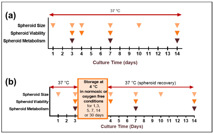
Cancer spheroids are very effective preclinical models to improve anticancer drug screening. In order to optimize and extend the use of spheroid models, these works were focused on the development of a new storage concept to maintain these models in the longer term using the Triple-Negative Breast Cancer MDA-MB-231 spheroid models. The results highlight that the combination of a temperature of 4 °C and oxygen-free conditions allowed the spheroid characteristics of OptiPASS® serum-free culture medium to preserve the spheroid characteristics during 3-, 5- or 7-day-long storage. Indeed, after storage they were returned to normal culture conditions, with recovered spheroids presenting similar growth rates (recovery = 96.2%), viability (Live/Dead® profiles) and metabolic activities (recovery = 90.4%) compared to nonstored control spheroids. Likewise, both recovered spheroids (after storage) and nonstored controls presented the same response profiles as two conventional drugs, i.e., epirubicin and cisplatin, and two anti-PARP1 targeted drugs-i.e., olaparib and veliparib. This new original storage concept seems to induce a temporary stop in spheroid growth while maintaining their principal characteristics for further use. In this way, this innovative and simple storage concept may instigate future biological sample preservation strategies.

Keywords: MDA-MB-231 spheroids; drug screening; new spheroid storage concept; preclinical spheroid models; triple-negative breast cancer.


Conflict of interest statement

The authors declare no conflict of interest.

Figures

Figure A1
Figure A1
Validation of the oxygen-free atmosphere generating system.
Figure 1
Figure 1
Schematic representation of MDA-MB-231 spheroids cold storage experiments. MDA-MB-231 spheroids were cultured in RPMI 1640 fetal calf serum-supplemented or OptiPASS® medium and maintained in classic cell culture conditions (humid incubator, 37 °C, 5% CO2). (a) For nonstored control condition, spheroids were continuously maintained at 37 °C, i.e., in classic culture conditions, for 14 days. (b) For storage experiments, after 3 days of culture, the spheroids in microplates were stored at 4 °C in normoxic or oxygen-free conditions for 1, 3, 5, 7, 14 or 30 days. After storage, spheroids in microplates were replaced in classic culture conditions for 10 supplemental days—i.e., between the 4th and the 14th days of culture.
Figure 2
Figure 2
MDA-MB-231 spheroid preservation analysis after 4 °C storage in RPMI1640 and OptiPASS® media culture. Three-day-old spheroids were placed for 1 or 3 days at 4 °C before being exposed to normal culture conditions—i.e., 37 °C, 5% CO2 in both RPMI1640 and OptiPASS® medium cultures. Spheroid integrity aspect and size (a) in RPMI and (e) in OptiPASS® were analyzed with bright field microscopy and object size algorithm (Cytation™3MV, Gen5, BioTek®-M = 4X, scale bar = 1000 µm). Spheroid cell proliferation was measured by the analysis of growth curve slopes between D4 and D14 (b) in RPMI and (f) in OptiPASS®, reflecting proliferation capacities and recovery of cell spheroids after storage step. Spheroid cell viability after storage was analyzed by Live/Dead® tests (c) in RPMI and (g) in OptiPASS® with green fluorescence for viable cells and red fluorescence for dead cells, using Cytation™3MV equipped with GFP and IP fluorescence cubes (M = 4X, scale bar = 1000 µm). Spheroid cell metabolic activity change after storage was quantified using the resazurin test (d) in RPMI and (h) in OptiPASS®, at D3, D7 and D14 with normalized 593 nm Fluorescence Intensity (FI) measures. For each storage condition, significances compared to nonstored condition were indicated as ns (not significant; p > 0.05), * p < 0.05, **** p < 0.0001, ***** p < 0.00001.
Figure 3
Figure 3
MDA-MB-231 spheroid preservation study after three-day storage at 4 °C and oxygen-free conditions in RPMI1640 and OptiPASS® media culture. Three-day-old spheroids cultured in normal culture conditions, i.e., 37 °C, 5% CO2 in RPMI1640 or OptiPASS® media culture, were exposed to oxygen-free conditions for 3 days at 4 °C. Then, the cultures were exposed again to normal culture conditions in which spheroid recovery was analyzed at day 3, day 4 and day 14. Spheroid integrity aspect and size (a) in RPMI and (d) in OptiPASS® were analyzed with bright field microscopy and object size algorithm (Cytation™3MV, Gen5, BioTek®, M = 4X, scale bar = 1000 µm). Spheroid cell viability after storage was studied by Live/Dead® tests (a) in RPMI and (d) in OptiPASS® with green fluorescence, for viable cells and red fluorescence, as well as for dead cells, using Cytation™3MV equipped with GFP and IP fluorescence cubes (M = 4X, scale bar = 1000 µm). Spheroid cell proliferation was measured by the growth curve slope analysis between D4 and D14 (b) in RPMI and (e) in OptiPASS®. Spheroid metabolic activity after storage (c) in RPMI and (f) in OptiPASS® medium culture was quantified using the resazurin tests. For each storage condition, significances compared to nonstored condition were indicated as ns (not significant; p > 0.05), ** p < 0.01, *** p < 0.001.
Figure 4
Figure 4
MDA-MB-231 spheroid preservation analysis after several days of storage in oxygen-free and 4 °C storage in OptiPASS® medium culture. Three-day-old spheroids cultured in normal culture conditions, i.e., 37 °C, 5% CO2 in OptiPASS® medium culture were exposed to oxygen-free conditions at 4 °C for 5, 7, 14 or 30 days. After each storage time, spheroid recovery was analyzed in comparison to nonstored control spheroids. Samples’ integrity aspects and sizes (a) were studied with bright field microscopy and object size algorithm (Cytation™3MV, Gen5, BioTek®, M = 4X, scale bar = 1000 µm). Growth curve slopes were calculated using spheroids’ size values between the 4th and the 14th days of culture, reflecting recovery of proliferation capacities after storage step (b). Spheroid viability after storage was assessed by Live/Dead® tests showing viable cells (green fluorescence) and dead cells (red fluorescence) using Cytation™3MV equipped with GFP and IP fluorescence cubes (M = 4X, scale bar = 1000 µm) (c). Complementary to this, spheroid metabolic activity was quantified using resazurin tests (d). Otherwise, spheroid tumoral proliferation gradient analysis was carried out by ki67 immunostaining at D4, D7 and D14 and imaged with Texas Red filter (M = 10X, scale bar = 200 µm) on Cytation™3MV instrument (BioTek®). Ki67-positive nuclei (in orange) were identified among other nuclei (in blue) with adapted algorithm on Gen5 software (BioTek®, pictures showed for nonstored controls and spheroids stored for 7 days) (e). Global ki67-expression was quantified and compared between nonstored controls and 3-, 5- and 7-day-long cold and oxygen-free storage conditions in OptiPASS® medium (f). Finally, spheroid hypoxia level was studied using ROS-ID® kit staining at D3, D4 and D14 (g,h). Spheroids were imaged with Cytation™3MV equipped with Texas Red fluorescence cube (M = 4X, scale bar = 500 µm) (g). Hypoxia level was quantified by acquired fluorescent signal intensity with Gen5 software (BioTek®) (h). For each storage condition, significances compared to nonstored condition were indicated as ns (not significant; p > 0.05), ** p < 0.01, **** p < 0.0001, ***** p < 0.00001.
Figure 5
Figure 5
Anticancer drug sensitivity evaluation on recovered MDA-MB-231 spheroids after 4 °C and oxygen-free storage in OptiPASS® medium. Three-day-old spheroids cultured in normal culture conditions, i.e., 37 °C, 5% CO2 in OptiPASS® medium culture, were stored at 4 °C in oxygen-free conditions for 3, 5 or 7 days. After returning to normal culture conditions for 2 days, spheroids were treated with epirubicin (0.1, 1 or 10 µM), cisplatin (0.1, 1 or 10 µM), olaparib (0.5, 5 or 50 µM) or veliparib (0.5, 5 or 50 µM) up to D10. For each condition, growth curve slopes during treatment were calculated for epirubicin (a), cisplatin (d), olaparib (g) and veliparib (j). Live/Dead® test profiles were imaged with plate reader Cytation™3 MV (GFP and IP filters-M = 4X) for epirubicin (b), cisplatin (e), olaparib (h) and veliparib (k). Metabolic activity rates were evaluated with resazurin test (Cytation™3MV-fluorimetry 593nm) for epirubicin (c), cisplatin (f), olaparib (i) and veliparib (l). ns = p > 0.05.

Similar articles

Cited by

References

    1. Bray F., Ferlay J., Soerjomataram I., Siegel R.L., Torre L.A., Jemal A. Global Cancer Statistics 2018: GLOBOCAN Estimates of Incidence and Mortality Worldwide for 36 Cancers in 185 Countries. CA Cancer J. Clin. 2018;68:394–424. doi: 10.3322/caac.21492. - DOI - PubMed
    1. Schmadeka R., Harmon B.E., Singh M. Triple-Negative Breast Carcinoma: Current and Emerging Concepts. Am. J. Clin. Pathol. 2014;141:462–477. doi: 10.1309/AJCPQN8GZ8SILKGN. - DOI - PubMed
    1. Palma G., Frasci G., Chirico A., Esposito E., Siani C., Saturnino C., Arra C., Ciliberto G., Giordano A., D’Aiuto M. Triple Negative Breast Cancer: Looking for the Missing Link between Biology and Treatments. Oncotarget. 2015;6:26560–26574. doi: 10.18632/oncotarget.5306. - DOI - PMC - PubMed
    1. Bryant H.E., Schultz N., Thomas H.D., Parker K.M., Flower D., Lopez E., Kyle S., Meuth M., Curtin N.J., Helleday T. Specific Killing of BRCA2-Deficient Tumours with Inhibitors of Poly(ADP-Ribose) Polymerase. Nature. 2005;434:913–917. doi: 10.1038/nature03443. - DOI - PubMed
    1. Lin P.-H., Chen M., Tsai L.-W., Lo C., Yen T.-C., Huang T.Y., Chen C.-K., Fan S.-C., Kuo S.-H., Huang C.-S. Using Next-Generation Sequencing to Redefine BRCAness in Triple-Negative Breast Cancer. Cancer Sci. 2020;111:1375–1384. doi: 10.1111/cas.14313. - DOI - PMC - PubMed

LinkOut - more resources