. 2021 Apr 14;13(8):1865.
doi: 10.3390/cancers13081865.

Tyrosine Kinase c-MET as Therapeutic Target for Radiosensitization of Head and Neck Squamous Cell Carcinomas

Affiliations

Tyrosine Kinase c-MET as Therapeutic Target for Radiosensitization of Head and Neck Squamous Cell Carcinomas

Lina Lüttich et al. Cancers (Basel). .

Abstract

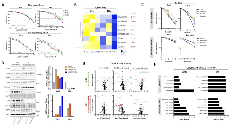
The receptor tyrosine kinase c-MET activates intracellular signaling and induces cell proliferation, epithelial-to-mesenchymal-transition and migration. Within the present study, we validated the prognostic value of c-MET in patients with head and neck squamous cell carcinoma (HNSCC) treated with radio(chemo)therapy using the Cancer Genome Atlas database and found an association of increased MET gene expression and protein phosphorylation with reduced disease-specific and progression-free survival. To investigate the role of c-MET-dependent radioresistance, c-MET-positive cells were purified from established HNSCC cell lines and a reduced radiosensitivity and enhanced sphere-forming potential, compared to the c-MET-depleted cell population, was found in two out of four analyzed cell lines pointing to regulatory heterogeneity. We showed that c-MET is dynamically regulated after irradiation in vitro and in vivo. Interestingly, no direct impact of c-MET on DNA damage repair was found. The therapeutic potential of eight c-MET targeting agents in combination with irradiation demonstrated variable response rates in six HNSCC cell lines. Amongst them, crizotinib, foretinib, and Pha665752 exhibited the strongest radiosensitizing effect. Kinase activity profiling showed an association of crizotinib resistance with compensatory PI3K/AKT and MAP kinase signaling. Overall, our results indicate that c-MET is conferring radioresistance in HNSCC through modulation of intracellular kinase signaling and stem-like features.

Keywords: c-MET kinase signaling; cancer stem cells; head and neck squamous cell carcinoma; radiotherapy; resistance.


Conflict of interest statement

M.B.: In the past five years, Michael Baumann received funding for his research projects and for educational grants to the University of Dresden by Bayer AG (2016–2018), Merck KGaA (2014–open) and Medipan GmbH (2014–2018). He is on the supervisory board of HI-STEM gGmbH (Heidelberg) for the German Cancer Research Center (DKFZ, Heidelberg) and also member of the supervisory body of the Charité University Hospital, Berlin. As former chair of OncoRay (Dresden) and present CEO and Scientific Chair of the German Cancer Research Center (DKFZ, Heidelberg), he has been or is responsible for collaborations with a multitude of companies and institutions, worldwide. In this capacity, he has discussed potential projects and signed contracts for research funding and/or collaborations with industry and academia for his institute(s) and staff, including but not limited to pharmaceutical companies such as Bayer, Boehringer Ingel-heim, Bosch, Roche and other companies such as Siemens, IBA, Varian, Elekta, Bruker, etc. In this role, he was/is also responsible for the commercial technology transfer activities of his institute(s), including the creation of start-ups and licensing. This includes the DKFZ-PSMA617 related patent portfolio [WO2015055318 (A1), ANTIGEN (PSMA)] and similar IP portfolios. Baumann confirms that, to the best of his knowledge, none of the above funding sources were involved in the preparation of this paper. M.K.: In the past five years, Krause received funding for her research projects by IBA (2016), Merck KGaA (2014–2018 for preclinical study; 2018–2020 for clinical study) and Medipan GmbH (2014–2018). She is involved in an ongoing publicly funded (German Federal Ministry of Education and Research) project with the companies Medipan, Attomol GmbH, GA Generic Assays GmbH, Gesellschaft für medizinische und wissenschaftliche genetische Analysen, Lipotype GmbH and PolyAn GmbH (2019–2021). For the present study, Dr. Krause confirms that none of the above-mentioned funding sources were involved. A.L.: Linge is involved in an ongoing publicly funded (German Federal Ministry of Education and Research) project with the companies Medipan, Attomol GmbH, GA Generic Assays GmbH, Gesellschaft für medizinische und wissenschaftliche genetische Analysen, Lipotype GmbH and PolyAn GmbH (2019–2021). For the present manuscript, Linge confirms that none of the above-mentioned funding sources were involved. The other authors declare no conflict of interest.

Figures

Figure 1
Figure 1
Identification of increased c-MET signaling in radioresistant head and neck squamous cell carcinoma (HNSCC) cell lines and patient data sets. (A) Schematic illustration of the generation of multiple irradiated (IR) sublines from established HNSCC cell lines. (B) MET gene expression analysis in parental compared to IR-sublines from FaDu and Cal33 based on Agilent array data (n = 3). (C) Comparative gene expression analysis of the MET regulatory gene set (31 genes, BioCarta, Human_RefSeq, ID: M19358) in Cal33- and FaDu-IR cells compared to parental control line demonstrated that 16 out of 31 genes are significantly altered (n = 3, p < 0.05). (D) The intersection between FaDu and Cal33 cell line for differential regulated genes (p < 0.05, fold change > 1.5, n = 39) in IR-sublines compared to parental control was analyzed for involvement in canonical pathways based on GSEA and g: Profiler webtool. (E) Kinase activity profiling (PamGene) identified altered intracellular serine/threonine kinases (STK) and protein tyrosine kinase (PTK) signaling in Cal33-IR cells compared to parental control (n = 3). Significantly increased kinase substrates involve, e.g., epidermal growth factor receptor (EGFR), cyclin-dependent kinase 1 (CDK1) and mesenchymal-epithelial transition factor/hepatocyte growth factor receptor (MET/HGFR) (fold change > 1.5, p value < 0.05). Upstream kinase prediction and pathway analysis including all significant regulated kinase substrates identified intracellular PI3K/AKT, Ras and EGFR signaling as key pathways altered in Cal33-IR clones. (F) Kaplan–Meier analyses validated the prognostic potential of MET gene expression for patient stratification and significantly predict disease-specific (p = 0.019, HR = 1.997, n = 443) and progression-free survival (p = 0.043, HR = 1.574, n = 474) in the TCGA-HNSCC data set. Subgroup analysis of patients with HNSCC treated with (n = 255) or without (n = 135) radiotherapy (RT) illustrated that patients with high MET expression show a significant reduction for disease-specific survival upon radiotherapy (p = 0.033, HR = 2.431). In HNSCC patients treated with surgery or chemotherapy alone MET expression does not have any prognostic potential (p = 0.677). (G) In silico analysis of The Cancer Genome Atlas (TCGA) RNASeq data set for patients with HNSCC (n = 517) showed significant negative correlation of MET expression with human papillomavirus (HPV) status and tumor grade (* p < 0.05, **** p < 0.0001).
Figure 2
Figure 2
Functional properties of the high c-MET-expressing population in HNSCC. (A) Western blot analysis for c-MET protein and correlation to tumor control doses 50% (TCD50) as indicator of tumor radiosensitivity. (B) Gene expression analysis of MET in Cal33, FaDu, SAS, and UT-SCC-5 cell lines derived from xenograft tumors that were further correlated to previously published tumor control dose 50% (TCD50) values [39,40,41]. (C) Proportions of c-MET positivity in different HNSCC cell lines were analyzed by flow cytometry. (D) Purification of c-MET high- and low-expressing cell populations (5%) using fluorescence-activated cell sorting (FACS). Reanalysis of the sorted populations demonstrated a purity of >99%. (E) The cell-intrinsic radiosensitivity of the purified c-MET-high- and low-expressing population was analyzed with a 3D-matrigel based colony-formation assay in 96-well plates. Dose-response curves illustrate the increased cell survival of c-MET+ cells in comparison to c-MET low-expressing cells upon irradiation with different doses (2, 4, 6, and 8 Gy) in SAS and UT-SCC-5 cell lines, while no differences were seen in FaDu and Cal33 (n = 3). (F) The DNA repair capacity of c-MET-high (c-MET+) and -low (c-MET) population upon 4 Gy irradiation was determined through the yH2AX-foci assay (n = 3, scale bar = 10 µm). The phosphorylation of histone 2AX indicates DNA double strand breaks and was investigated using immunofluorescence analysis 30 min (initial foci) and 24 h (residual foci) after 4 Gy irradiation (* p < 0.05). (G) In silico analysis of the TCGA data set correlating the MET expression in patients with HNSCC (n = 517) with the occurrence of canonical pathways (Molecular Signatures Database (MSigDB), Broad Institute) identified a positive correlation with cell motility, receptor tyrosine kinase (RTK) signaling and focal adhesion, while signaling pathways involved in DNA repair, amino acid metabolism and DNA damage showed significantly negative correlation. (H) Chemical inhibition of c-MET with crizotinib (10 µM), mTOR/PI3K with BEZ235, Chk1 with LY2880070, and WNT blockade with XAV 939 (10 nM) for 3 days influenced the percentage of c-MET and CD44-positive population in Cal33, FaDu, and UT-SCC-5. (I) MET gene expression in primary tumors decreases significantly with increasing nodal status (* p < 0.05, TCGA) and is significantly down-regulated in primary tumors of patients with HNSCC with both, increasing nodal (N) status and distant metastasis (* p < 0.05, ** p < 0.01, **** p < 0.0001, ns (not significant)).
Figure 3
Figure 3
Cellular plasticity and self-renewal properties of the c-MET-population in HNSCC. (A) c-MET protein expression within Cal33, FaDu, and UT-SCC-5 xenograft tumors that were treated with 10 fractions of 2 Gy within two weeks. Correlation of tumor control doses 50% with ratio of c-MET protein in irradiated cohort to sham control. (B) Immunofluorescence-based evaluation of c-MET expression in s.c. xenograft tumors originating from Cal33, FaDu, and SAS that were treated with cisplatin-based radio/chemotherapy in 10 fractions after randomization when tumors reached a diameter of 7 mm (n = 8–15). Tumors were fixed 24 h after last fraction and the formalin-fixed paraffin-embedded (FFPE) tissue was cut into 4 µm sections and stained with anti-c-MET antibody. (C) Single dose irradiation with 4 Gy modulates the c-MET protein expression in Cal33, FaDu, and SAS differently in a time-dependent manner within one week. Semi-quantitative analysis of this Western blot using ImageJ demonstrated a dynamic up-regulation of c-MET in Cal33 and SAS as well as a down-regulation in FaDu cells (n = 2). (D) Flow cytometry-based analysis validated the irradiation-induced upregulation of c-MET on cell membrane 2 days after irradiation with 4 Gy. The depicted overlay histogram compares sham control (red) and post-irradiated cells (blue) for Cal33 and FaDu labeled with c-MET-Alexa488 antibody. (E) Immunofluorescence analysis illustrates an increased c-MET expression 5 days upon 4 Gy irradiation and a reduced c-MET signal in IR subclones. c-MET membrane expression and intracellular localization was visualized using an anti-c-MET-specific primary antibody recognized by an AlexaFluor488-labeled secondary antibody (green). The nuclei were stained with 4′,6-diamidino-2-phenylindole (DAPI, blue, scale bar = 50 μm). Quantification of c-Met expression per cell was performed using ImageJ analysis through measurement of mean pixel intensity. (F) Cell conversion and plasticity of FACS-purified c-MET-high and low populations in Cal33 and SAS was analyzed 7 days after sorting and seeding of 27,000 cells per well within 6-well-plates by flow cytometry; 100% pure Cal33 c-MET+ culture reduces c-MET+ proportion to 81.7% after 7 days in culture, while pure c-MET culture increases c-MET+ population to 30.3%. SAS cell line showed to be more plastic than Cal33 and switches back to nearly original proportion of 90% c-MET+ cells within 7 days. (G) FACS-purified c-MET-high and low population from SAS and UT-SCC-5 cell line were plated under non-adhesive, single cell conditions into low-attachment plates with growth factor defined mammary epithelial basal medium (MEBM). Plates were scanned 14 days after plating with imaging cytometry and formed spheres with a size >100 µm were counted manually. ImageJ was used to calculate sphere-forming potential for each population indicative for self-renewal and stem-like capacity (n = 3, * p < 0.05). (H) Co-expression of c-MET with other cancer stem cells (CSC) and radioresistance markers such as CD44, CD98, and EGFR was analyzed using multicolor flow cytometry (BD FACS Celesta) and illustrates about 50% co-expression in the tested cell lines Cal33, SAS, and Detroit562. (I) In silico analysis of TCGA RNASeq data set for patients with HNSCC (n = 517) demonstrated significantly, positive correlation of MET gene expression with CD44, SLC3A2, and EGFR (Pearson correlation) (* p < 0.05, ** p < 0.01).
Figure 4
Figure 4
Cellular effects of siRNA-mediated MET-specific gene knock-down in HNSCC cell lines. (A) Validation of down-regulated c-MET protein expression 24 h upon transfection with 50 pmol of c-MET specific siRNA#1 and #2 in comparison to unspecific scrambled control (scr). Western blot analysis demonstrated highly efficient c-MET (175 kDa) down-regulation in Cal33 and FaDu as well as their irradiated sublines (IR). Glyceraldehyde 3-phosphate dehydrogenase (GAPDH, 35 kDa) was used as internal loading control (n = 2). (B,C) Clonogenic survival and plating efficiency (b) in standard 2D-colony-assay 10 days after c-MET knock-down for Cal33 and FaDu cell line as well as their irradiated sub-lines (IR) in comparison to scramble (scr) control (n = 3, * p < 0.05, mean ± SEM). Linear-quadratic model of cell survival curves after irradiation with 0, 2, 4, or 6 Gy (c) (n = 3, * p < 0.05, mean ± SEM). The number of colonies within 10 days is calculated to plated cell number and depicted as plating efficiency. The surviving fraction illustrates the clonogenic survival depending on the irradiation dose and is normalized to plating efficiency. (D) Western blot analysis of Detroit562 cells 24 h after c-MET knock-down validated the effects on c-MET proteins without affecting c-MET phosphorylation and other downstream kinases such as AKT and ERK1/2. β-actin was used as loading control. (E) Clonogenic survival of metastasis cell line Detroit562 which originates from pleural effusion (primary origin: pharynx) 10 days after MET knock-down upon transfection with 30 pmol of c-MET specific siRNA#1 or #2 in 3D-matrigel-based cultures treated with increasing doses of irradiation (2, 4, 6, and 8 Gy) (n = 3, mean ± SEM). (F) In silico analysis of TCGA-HNSCC proteome database (n = 162) for the phosphorylated c-MET (Y1235, pMET) protein analyzed via reverse phase protein array illustrates patient stratification with Kaplan–Meier curves. HNSCC patients with high level of pMET have a significantly lower disease-specific survival in comparison to patients with low pMET level in local tumor biopsies (p = 0.006, HR = 2.433). Subgroup analysis for HNSCC patients treated with radiotherapy (RT, n = 65) does not show significant differences (p = 0.309). (G) Correlation analysis of MET gene expression with phosphoprotein analysis illustrates no association within the HNSCC-TCGA data set (r = 0.089, Spearman) (* p < 0.05, ** p < 0.01, *** p < 0.001).
Figure 5
Figure 5
c-MET-specific chemical targeting to induce intracellular sensitization of HNSCC cell lines to ionizing radiation. (A) The half maximal inhibitory concentrations (IC50) of eight different c-MET-targeting agents were determined on different time points (24 h, 48 h, and 72 h) with increasing compound concentrations (1–100 µM) and in combination with irradiation (4 Gy) using cell viability assay determining intracellular adenosine triphosphate (ATP) level (CellTiter-Glo, Promega) (n = 2). (B) Unsupervised cluster analysis illustrates radiosensitizing potential of eight c-MET-targeting chemical compounds in Cal33, FaDu, and SAS cell line. (C) Three compounds with significant radiosensitization in cell survival assay were validated within a 3D-colony formation assay including 4 h inhibitory treatment before irradiation with 2, 4, 6, and 8 Gy of X-rays. The number of colonies was examined after 10 days of culture. Depicted are the surviving fractions normalized to plating efficiency of sham control (n = 3, mean ± SEM, * p < 0.05). (D) Activity of different intracellular kinase signaling after 4 h and 24 h of treatment with crizotinib or Pha665752 within the cMET-targeting responsive cell line Cal33 and non-responding line SAS was analyzed by determining specific phosphorylation signals with Western blot (n = 2). (E) Kinome activity of responding cell line Cal33 and non-responding line SAS was analyzed using capture kinase assay (PTK and STK PamChip) after 4 h treatment with crizotinib (IC10), 24 h after 4 Gy and crizotinib combined with irradiation in comparison to control (sham and DMSO) (n = 3). Venn diagrams illustrate identified peptides significantly altered within the treatment group (p < 0.05). (F) All significantly phosphorylated peptides within a certain treatment group were included to perform pathway analysis with the Molecular Signatures Database (MSigDB) to illustrate altered intracellular upstream protein kinase signaling pathways (* p < 0.05).

Similar articles

Cited by

References

    1. Rettig E.M., D’Souza G. Epidemiology of Head and Neck Cancer. Surg. Oncol. Clin. N. Am. 2015;24:379–396. doi: 10.1016/j.soc.2015.03.001. - DOI - PubMed
    1. Sung H., Ferlay J., Siegel R.L., Laversanne M., Soerjomataram I., Jemal A., Bray F. Global Cancer Statistics 2020: GLOBOCAN Estimates of Incidence and Mortality Worldwide for 36 Cancers in 185 Countries. CA A Cancer J. Clin. 2021 doi: 10.3322/caac.21660. - DOI - PubMed
    1. Gatta G., Botta L., Sánchez M.J., Anderson L.A., Pierannunzio D., Licitra L., EUROCARE Working Group Prognoses and Improvement for Head and Neck Cancers Diagnosed in Europe in Early 2000s: The EUROCARE-5 Population-Based Study. Eur. J. Cancer. 2015;51:2130–2143. doi: 10.1016/j.ejca.2015.07.043. - DOI - PubMed
    1. Lee N., Puri D.R., Blanco A.I., Chao K.S.C. Intensity-Modulated Radiation Therapy in Head and Neck Cancers: An Update. Head Neck. 2007;29:387–400. doi: 10.1002/hed.20332. - DOI - PubMed
    1. Argiris A., Li S., Savvides P., Ohr J.P., Gilbert J., Levine M.A., Chakravarti A., Haigentz M., Jr., Saba N.F., Ikpeazu C.V., et al. Phase III Randomized Trial of Chemotherapy with or without Bevacizumab in Patients with Recurrent or Metastatic Head and Neck Cancer. J. Clin. Oncol. 2019;37:3266–3274. doi: 10.1200/JCO.19.00555. - DOI - PMC - PubMed

LinkOut - more resources