. 2021 Apr 14;22(8):4030.
doi: 10.3390/ijms22084030.

Impaired Ca2+ Sensitivity of a Novel GCAP1 Variant Causes Cone Dystrophy and Leads to Abnormal Synaptic Transmission Between Photoreceptors and Bipolar Cells

Affiliations

Impaired Ca2+ Sensitivity of a Novel GCAP1 Variant Causes Cone Dystrophy and Leads to Abnormal Synaptic Transmission Between Photoreceptors and Bipolar Cells

Valerio Marino et al. Int J Mol Sci. .

Abstract

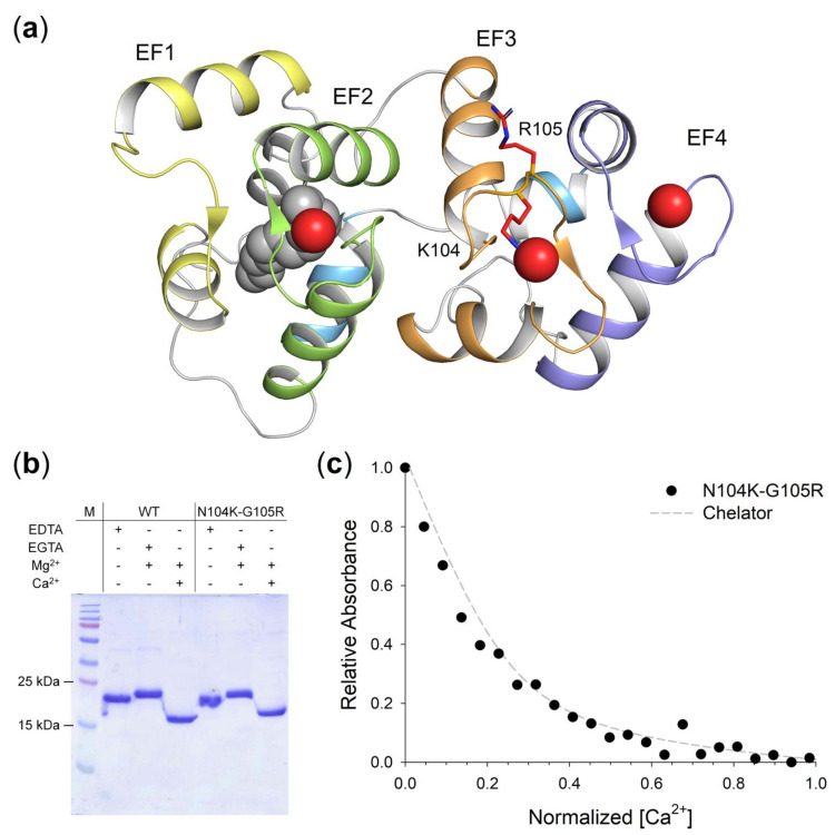
Guanylate cyclase-activating protein 1 (GCAP1) is involved in the shutdown of the phototransduction cascade by regulating the enzymatic activity of retinal guanylate cyclase via a Ca2+/cGMP negative feedback. While the phototransduction-associated role of GCAP1 in the photoreceptor outer segment is widely established, its implication in synaptic transmission to downstream neurons remains to be clarified. Here, we present clinical and biochemical data on a novel isolate GCAP1 variant leading to a double amino acid substitution (p.N104K and p.G105R) and associated with cone dystrophy (COD) with an unusual phenotype. Severe alterations of the electroretinogram were observed under both scotopic and photopic conditions, with a negative pattern and abnormally attenuated b-wave component. The biochemical and biophysical analysis of the heterologously expressed N104K-G105R variant corroborated by molecular dynamics simulations highlighted a severely compromised Ca2+-sensitivity, accompanied by minor structural and stability alterations. Such differences reflected on the dysregulation of both guanylate cyclase isoforms (RetGC1 and RetGC2), resulting in the constitutive activation of both enzymes at physiological levels of Ca2+. As observed with other GCAP1-associated COD, perturbation of the homeostasis of Ca2+ and cGMP may lead to the toxic accumulation of second messengers, ultimately triggering cell death. However, the abnormal electroretinogram recorded in this patient also suggested that the dysregulation of the GCAP1-cyclase complex further propagates to the synaptic terminal, thereby altering the ON-pathway related to the b-wave generation. In conclusion, the pathological phenotype may rise from a combination of second messengers' accumulation and dysfunctional synaptic communication with bipolar cells, whose molecular mechanisms remain to be clarified.

Keywords: GUCA1A; bipolar cells; calcium binding proteins; cone dystrophy; guanylate cyclase; neuronal calcium sensor; photoreceptors; phototransduction; retinal degeneration; synaptic transmission.


Conflict of interest statement

The authors declare no conflict of interests.

Figures

Figure 1
Figure 1
Fundus autofluorescence and optical coherence tomography recorded in the study patient (top rows) and in a healthy control (bottom rows). Green arrows represent the direction of OCT scan. Note the large area of atrophy in the central retina and the thinning of the outer nuclear layer. Scale bars = 200 µm.
Figure 2
Figure 2
Standard electroretinography recorded in a control subject (a 48-year-old female, left columns) and in the study patient (right columns). Note that cone ERGs were unrecordable in the patient. Rod ERGs showed reduced b-wave amplitude and a lower-than-normal b/a wave amplitude ratio in the dark-adapted mixed rod-cone ERG.
Figure 3
Figure 3
Regulation of guanylate cyclases by GCAP1 variants. Enzymatic regulation of (a) human GC1 and (b) human GC2 by 5 µM GCAP1 WT and N104K-G105R variants in activating (low Ca2+, <19 nM) and inhibiting conditions (high Ca2+, ~30 µM). Presented data are an average ± SD of 3 independent experiments.
Figure 4
Figure 4
Structural model of N104K-G105R GCAP1 and assessment of Ca2+-affinity of GCAP1 variants. (a) 3D structural model of human N104K-G105R GCAP1 in the Ca2+-loaded form. Protein structure shown in cartoon form, with structural regions specifically colored (N-terminal helix grey, EF1 yellow, EF2 green, EF3 orange, EF4 blue, C-terminal helices cyan). Myristoyl moiety is represented as grey spheres, Ca2+ ions as red spheres, residues K104 and R105 as red sticks, with N atoms in blue. (b) 15% SDS-PAGE of 20 μM GCAP1 WT and N104K-G105R variants in the presence of 5 mM EDTA, 5 mM EGTA + 1.1 mM Mg2+ or 1 mM Mg2+ + 5 mM Ca2+. (c) Example of Ca2+ titration curve for N104K-G105R GCAP1. Normalized absorption of 5,5′Br2-BAPTA upon Ca2+-binding in competition with N104K-G105R GCAP1 (black circles) in the presence of 1 mM Mg2+, together with the theoretical simulated curve of the chelator in the absence of competition (grey dashed line). Ca2+-concentration considers the dilution upon Ca2+-addition, normalization details are reported in the Methods section.
Figure 5
Figure 5
Quaternary structure and aggregation propensity of N104K-G105R monitored by analytical SEC and DLS. (a) Analytical SEC chromatograms of 42 µM N104K-G105R in the presence of 500 μM EGTA + 1 mM Mg2+ (blue) or 500 μM Ca2+ + 1 mM Mg2+ (red). (b) DLS measurements of 42 µM N104K-G105R in the presence of 500 μM EGTA + 1 mM Mg2+ (blue) or 500 μM Ca2+ + 1 mM Mg2+ (red). (c) Time evolution of the mean count rate over 15 h of 42 µM N104K-G105R in the presence of 500 μM EGTA + 1 mM Mg2+ (blue) or 500 μM Ca2+ + 1 mM Mg2+ (red). Analytical SEC and DLS experiments were carried out in 20 mM Tris-HCl pH 7.5, 150 mM KCl, 1 mM DTT buffer. Estimations of the apparent MM and the hydrodynamic diameter are reported in Table 2.
Figure 6
Figure 6
Structural and stability changes of N104K-G105R upon ion binding assessed by Circular Dichroism (CD) spectroscopy. (a) Far UV CD spectra of 10 μM N104K-G105R in the presence of 300 μM EGTA (black) and after serial additions of 1 mM Mg2+ (blue) and 600 μM Ca2+ (red). (b) Near UV CD spectra of 42 μM N104K-G105R in the presence of 500 μM EGTA (black) and after serial additions of 1 mM Mg2+ (blue) and 1 mM Ca2+ (red). (c) Thermal denaturation profiles of 10 μM N104K-G105R in the presence of 300 μM EGTA (black), 300 μM EGTA + 1 mM Mg2+ (blue) and 1 mM Mg2+ + 300 μM free Ca2+ (red). CD spectroscopy measurements were conducted in 20 mM Tris-HCl pH 7.5, 150 mM KCl, 1 mM DTT buffer. Thermal denaturation profiles were collected by monitoring the ellipticity at 222 nm between 20 and 96 °C and were fitted to a function accounting for thermodynamic contributions described in the Methods section.
Figure 7
Figure 7
Cα-root-mean-square fluctuation (RMSF) projected on the 3D structure of GCAP1 WT (upper panels) and N104K-G105R (lower panels) in their activating (Mg2+-loaded, left) and inhibiting (Ca2+-loaded, right) states. Protein structure is displayed as a tube cartoon with diameter proportional to the RMSF; Mg2+ and Ca2+ ions are shown as spheres. Structures are colored in a rainbow scheme representing RMSF values from 0.6 to 3 Å (see Figure S4 for Cα-RMSF profiles).

Similar articles

Cited by

References

    1. Koch K.W., Dell’Orco D. A calcium-relay mechanism in vertebrate phototransduction. ACS Chem. Neurosci. 2013;4:909–917. doi: 10.1021/cn400027z. - DOI - PMC - PubMed
    1. Koch K.W., Dell’Orco D. Protein and signaling networks in vertebrate photoreceptor cells. Front. Mol. Neurosci. 2015;8:67. doi: 10.3389/fnmol.2015.00067. - DOI - PMC - PubMed
    1. Korenbrot J.I. Speed, sensitivity, and stability of the light response in rod and cone photoreceptors: Facts and models. Prog. Retin. Eye Res. 2012;31:442–466. doi: 10.1016/j.preteyeres.2012.05.002. - DOI - PMC - PubMed
    1. Peshenko I.V., Dizhoor A.M. Guanylyl cyclase-activating proteins (GCAPs) are Ca2+/Mg2+ sensors: Implications for photoreceptor guanylyl cyclase (RetGC) regulation in mammalian photoreceptors. J. Biol. Chem. 2004;279:16903–16906. doi: 10.1074/jbc.C400065200. - DOI - PubMed
    1. Peshenko I.V., Dizhoor A.M. Ca2+ and Mg2+ binding properties of GCAP-1. Evidence that Mg2+-bound form is the physiological activator of photoreceptor guanylyl cyclase. J. Biol. Chem. 2006;281:23830–23841. doi: 10.1074/jbc.M600257200. - DOI - PubMed

MeSH terms

LinkOut - more resources