Review
. 2021 Apr 14;22(8):4053.
doi: 10.3390/ijms22084053.

Quantification of Dendritic Spines Remodeling under Physiological Stimuli and in Pathological Conditions

Affiliations
Review

Quantification of Dendritic Spines Remodeling under Physiological Stimuli and in Pathological Conditions

Ewa Bączyńska et al. Int J Mol Sci. .

Abstract

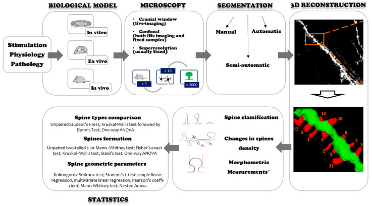
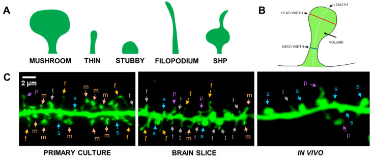
Numerous brain diseases are associated with abnormalities in morphology and density of dendritic spines, small membranous protrusions whose structural geometry correlates with the strength of synaptic connections. Thus, the quantitative analysis of dendritic spines remodeling in microscopic images is one of the key elements towards understanding mechanisms of structural neuronal plasticity and bases of brain pathology. In the following article, we review experimental approaches designed to assess quantitative features of dendritic spines under physiological stimuli and in pathological conditions. We compare various methodological pipelines of biological models, sample preparation, data analysis, image acquisition, sample size, and statistical analysis. The methodology and results of relevant experiments are systematically summarized in a tabular form. In particular, we focus on quantitative data regarding the number of animals, cells, dendritic spines, types of studied parameters, size of observed changes, and their statistical significance.

Keywords: dendritic spine analysis; dendritic spine morphology; dendritic spines; neuronal remodeling; spine remodeling; structural plasticity; synaptic plasticity.


Conflict of interest statement

The authors declare no conflict of interest.

Figures

Figure 1
Figure 1
Typical experimental workflow. Green color indicates the dendrite, red color indicates the dendritic spines.
Figure 2
Figure 2
Morphological diversity of dendritic spines. (A) Spine shape classification: mushroom (m), thin (t), stubby (s), filopodium (f), SHP-spine head protrusion (p); (B) definition of morphometric parameters; (C) microscopic images of dendrites covered with dendritic spines obtained from in vitro (primary culture), ex vivo (brain slice), and in vivo (cranial window) imaging. Scale bar: 2 μm.
Figure 3
Figure 3
Different imaging modalities; fluorescent image of a dendrite with spines. Different types of neurons and staining techniques were used. The actual colors were changed in postprocessing to the green. (A) Live-cell imaging of primary hippocampal culture transfected with plasmid-encoded RFP (B) Confocal image combined with Airyscan processing of hippocampal fixed slices biolistically labeled with DiI staining (C) transcranial two-photon imaging of the motor cortex in a freely moving mouse. (D) Confocal image of organotypic hippocampal slice culture biolistically transfected with plasmid-encoded RFP. (E) Confocal image of fixed primary hippocampal culture transfected with plasmid-encoded GFP, (F) Confocal image of thick hippocampal brain slice marked with DiI. Scale bar: 2 μm.

References

    1. Kasai H., Fukuda M., Watanabe S., Hayashi-Takagi A., Noguchi J. Structural dynamics of dendritic spines in memory and cognition. Trends Neurosci. 2010;33:121–129. doi: 10.1016/j.tins.2010.01.001. - DOI - PubMed
    1. Bola M., Borchardt V. Cognitive Processing Involves Dynamic Reorganization of the Whole-Brain Network’s Functional Community Structure. J. Neurosci. 2016;36:3633–3635. doi: 10.1523/JNEUROSCI.0106-16.2016. - DOI - PMC - PubMed
    1. Penzes P., Cahill M.E., Jones K.A., VanLeeuwen J.-E., Woolfrey K.M. Dendritic spine pathology in neuropsychiatric disorders. Nat. Neurosci. 2011;14:285–293. doi: 10.1038/nn.2741. - DOI - PMC - PubMed
    1. Bailey C.H., Kandel E.R., Harris K.M. Structural Components of Synaptic Plasticity and Memory Consolidation. Cold Spring Harb. Perspect. Biol. 2015;7:a021758. doi: 10.1101/cshperspect.a021758. - DOI - PMC - PubMed
    1. Holtmaat A., Svoboda K. Experience-dependent structural synaptic plasticity in the mammalian brain. Nat. Rev. Neurosci. 2009;10:647–658. doi: 10.1038/nrn2699. - DOI - PubMed

MeSH terms

LinkOut - more resources