. 2021 Apr 14;22(8):4056.
doi: 10.3390/ijms22084056.

Protective Effect of Quercetin on Sodium Iodate-Induced Retinal Apoptosis through the Reactive Oxygen Species-Mediated Mitochondrion-Dependent Pathway

Affiliations

Protective Effect of Quercetin on Sodium Iodate-Induced Retinal Apoptosis through the Reactive Oxygen Species-Mediated Mitochondrion-Dependent Pathway

Yuan-Yen Chang et al. Int J Mol Sci. .

Abstract

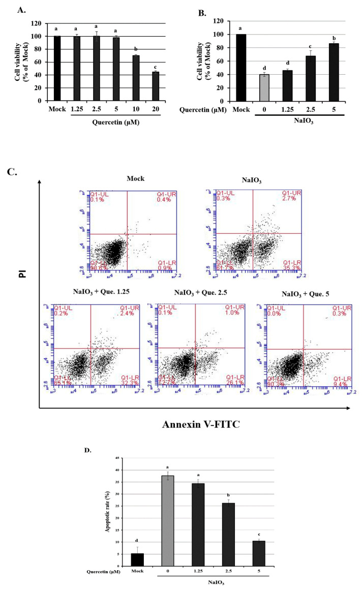
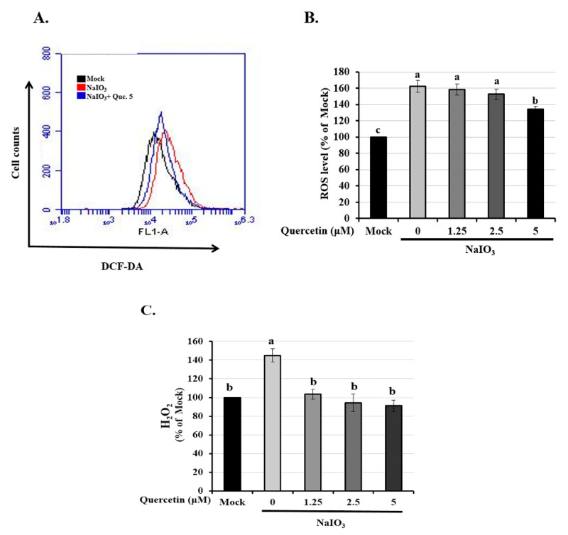
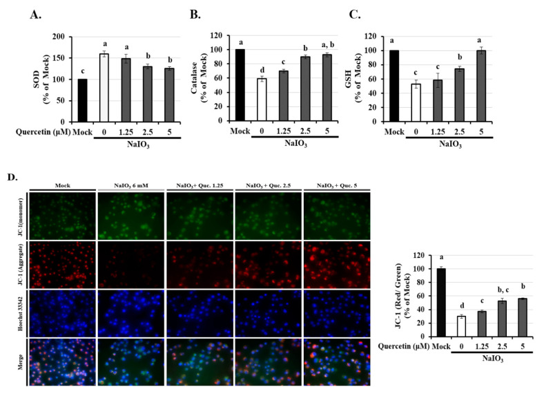
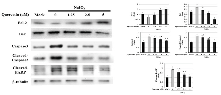
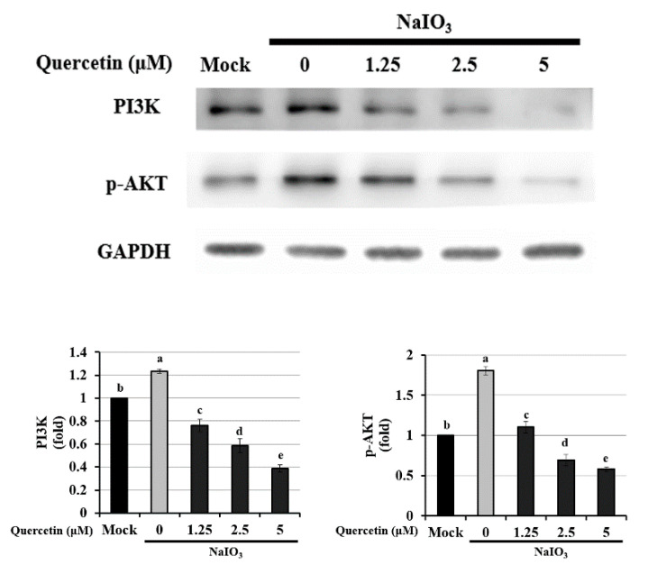
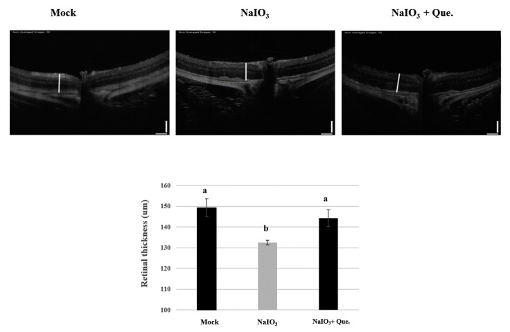
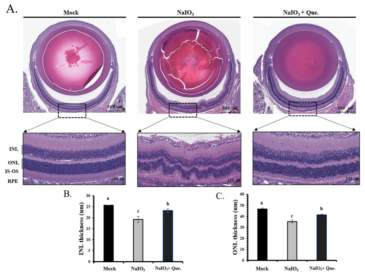
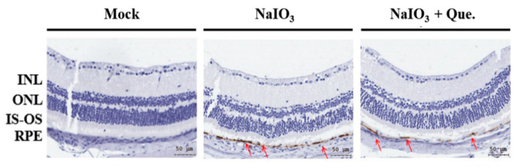
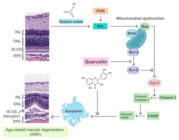
Age-related macular degeneration (AMD) leads to gradual central vision loss and is the third leading cause of irreversible blindness worldwide. The underlying mechanisms for this progressive neurodegenerative disease remain unclear and there is currently no preventive treatment for dry AMD. Sodium iodate (NaIO3) has been reported to induce AMD-like retinal pathology in mice. We established a mouse model for AMD to evaluate the effects of quercetin on NaIO3-induced retinal apoptosis, and to investigate the pertinent underlying mechanisms. Our in vitro results indicated that quercetin protected human retinal pigment epithelium (ARPE-19) cells from NaIO3-induced apoptosis by inhibiting reactive oxygen species production and loss of mitochondrial membrane potential as detected by Annexin V-FITC/PI flow cytometry. We also evaluated the relative expression of proteins in the apoptosis pathway. Quercetin downregulated the protein expressions of Bax, cleaved caspase-3, and cleaved PARP and upregulated the expression of Bcl-2 through reduced PI3K and pAKT expressions. Furthermore, our in vivo results indicated that quercetin improved retinal deformation and increased the thickness of both the outer nuclear layer and inner nuclear layer, whereas the expression of caspase-3 was inhibited. Taken together, these results demonstrate that quercetin could protect retinal pigment epithelium and the retina from NaIO3-induced cell apoptosis via reactive oxygen species-mediated mitochondrial dysfunction, involving the PI3K/AKT signaling pathway. This suggests that quercetin has the potential to prevent and delay AMD and other retinal diseases involving NaIO3-mediated apoptosis.

Keywords: age-related macular degeneration; apoptosis; human retinal pigment epithelium; mitochondrial membrane potential; quercetin; sodium iodate.


Conflict of interest statement

The authors declare no conflict of interest.

Figures

Figure 1
Figure 1
Cell viability of ARPE-19 cells treated with quercetin only or quercetin plus NaIO3. (A) ARPE-19 cells were treated with various concentrations of quercetin (1.25, 2.5 and 5 μM) for 24 h, and the cell viability was measured using a CCK-8 assay. (B) ARPE-19 cells were pretreated with various concentrations of quercetin (1.25, 2.5 and 5 μM) for 1.5 h and then with NaIO3 (6 mM) for 24 h, before cell viability was measured using the CCK-8 assay. (C) ARPE-19 cells were pretreated with various concentrations (1.25, 2.5 and 5 μM) of quercetin for 1.5 h before treatment with NaIO3 (6 mM) for 15 h. ARPE-19 cells were then detected by flow cytometry after staining with both Annexin V-FITC and PI for 30 min. (D) The percentage of apoptotic cells in each treatment group was quantified. Values represent the mean ± SD (n = 3). Data bars without letters in common (a–d) indicate a significant difference (p < 0.05).
Figure 2
Figure 2
Effect of quercetin on NaIO3-mediated ROS generation in ARPE-19 cells. ARPE-19 cells were pretreated with different concentrations of quercetin (1.25, 2.5, and 5 μM) for 1.5 h and then treated with NaIO3 (6 mM) for 15 h. (A,B) Cells were labeled with the fluorescent probe, 2′,7′ dichlorodihydrofluorescein diacetate, the intracellular ROS levels were quantitatively analyzed and the mean fluorescence intensity was calculated using flow cytometry. (C) ARPE-19 cells were pretreated with different concentrations of quercetin (1.25, 2.5, and 5 μM) for 1.5 h and then treated with NaIO3 (6 mM) for 24 h. The amount of H2O2 were measured by commercial assay kits. Values represent the mean ± SD (n = 3). Data bars without letters in common (a–c) indicate a significant difference (p < 0.05).
Figure 3
Figure 3
Effects of quercetin on antioxidant activity and mitochondrial dysfunction in ARPE-19 cells. ARPE-19 cells were pretreated with different concentrations of quercetin (1.25, 2.5, and 5 μM) for 1.5 h and then treated with NaIO3 (6 mM) for 15 h. The levels of (A) superoxide (SOD), (B) catalase, and (C) glutathione (GSH) were quantitatively analyzed, and the mean fluorescence intensity was calculated using flow cytometry. (D) Mitochondrial membrane potential was measured using the fluorescent probe JC-1. Loss of mitochondrial membrane depolarization (ΔΨm) was demonstrated by the change in JC-1 fluorescence from red (JC-1 aggregates) to green (JC-1 monomers). The fluorescent intensity ratio of JC-1 aggregates to monomers in treated cells. Values represent the mean ± SD (n = 3). Data bars without letters in common (a–d) indicate a significant difference (p < 0.05).
Figure 4
Figure 4
Western blotting was performed to assess the effects of quercetin on the expression of Bcl-2, Bax, caspase-3, cleaved caspase-3, and cleaved PARP in NaIO3-treated ARPE-19 cells. ARPE-19 cells were pretreated with different concentrations of quercetin (1.25, 2.5, and 5 μM) for 1.5 h and then treated with NaIO3 (6 mM) for 24 h. Data were normalized to β-tubulin. Values represent the mean ± SD (n = 3). Data bars without letters in common (a–d) indicate a significant difference (p < 0.05).
Figure 5
Figure 5
Western blotting to determine the expression of PI3K and p-AKT in NaIO3-treated ARPE-19 cells. ARPE-19 cells were pretreated with different concentrations of quercetin (1.25, 2.5, and 5 μM) for 1.5 h and then treated with NaIO3 (6 mM) for 24 h. Data were normalized to GAPDH. Values represent mean ± SD (n = 3). Data bars without letters in common (a–e) indicate a significant difference (p < 0.05).
Figure 6
Figure 6
Preventive effects of quercetin on the retinal thickness of NaIO3-treated mice. Optical coherence tomography (OCT) was performed 7 days after NaIO3 treatment for all three study groups. The retinal degenerative changes are shown. Vertical bar = 100 μm and horizontal bar = 120 μm; white line represents retinal thickness. Values represent mean ± SD (n = 6). Data bars without letters in common (a,b) indicate a significant difference (p < 0.05).
Figure 7
Figure 7
Protective effects of quercetin on retinal degeneration in NaIO3-treated mice. (A) Representative retinal sections (H&E staining) for the three groups from between 600 μm and 900 μm from the optic nerve along the superior and inferior hemiretina. ONL, outer nuclear layer; INL, inner nuclear layer; IS-OS, inner and outer segment of photoreceptor; RPE, retinal pigment epithelium. Scale bar = 50 μm. (B) INL and (C) ONL thickness were measured at six locations and the values were then averaged. Values represent the mean ± SD (n = 6). Data bars without letters in common (a–c) indicate a significant difference (p < 0.05).
Figure 8
Figure 8
Effect of quercetin on NaIO3-induced retinal apoptosis in mice. After 7 days of NaIO3 treatment, immunohistochemical staining was performed for cleaved caspase-3 in all three study groups. The red arrows indicate cleaved caspase-3 in the retinal pigment epithelial (RPE) layers. The vehicle-treated NaIO3 showed a significantly higher expression of cleaved caspase-3, while the mock and experimental groups showed significantly lower expression of cleaved caspase-3. Cleaved caspase-3 is shown in brown, and the red arrows are within the position of the RPE layer.
Figure 9
Figure 9
Illustration of the mechanism of NaIO3-induced retinal injury and the effects of quercetin. NaIO3 induces mitochondrial dysfunction through the PI3K/AKT signaling pathway and then Bax activates Cytochrome C, which causes apoptosis. Finally, IS-OS disruption occurs, which mimics AMD changes within the retinal tissue. Quercetin can activate Bcl-2 and inhibit apoptosis, thereby reducing the accumulation of oxidative damage.

References

    1. Gibbons A., Ali T.K., Waren D.P., Donaldson K.E. Causes and correction of dissatisfaction after implantation of presbyopia-correcting intraocular lenses. Clin. Ophthalmol. 2016;10:1965–1970. doi: 10.2147/OPTH.S114890. - DOI - PMC - PubMed
    1. Ambati J., Atkinson J.P., Gelfand B.D. Immunology of age-related macular degeneration. Nat. Rev. Immunol. 2013;13:438–451. doi: 10.1038/nri3459. - DOI - PMC - PubMed
    1. Klein R., Myers C.E., Buitendijk G.H., Rochtchina E., Gao X., de Jong P.T., Sivakumaran T.A., Burlutsky G., McKean-Cowdin R., Hofman A., et al. Lipids, lipid genes, and incident age-related macular degeneration: The three continent age-related macular degeneration consortium. Am. J. Ophthalmol. 2014;158:513–524.e3. doi: 10.1016/j.ajo.2014.05.027. - DOI - PMC - PubMed
    1. Mahr M.A., Hodge D.O., Erie J.C. Racial Differences in Age-Related Macular Degeneration and Associated Anti-Vascular Endothelial Growth Factor Intravitreal Injections among Medicare Beneficiaries. Ophthalmol. Retin. 2018;2:1188–1195. doi: 10.1016/j.oret.2018.05.005. - DOI - PubMed
    1. Rozing M.P., Durhuus J.A., Krogh Nielsen M., Subhi Y., Kirkwood T.B., Westendorp R.G., Sorensen T.L. Age-related macular degeneration: A two-level model hypothesis. Prog. Retin. Eye Res. 2020;76:100825. doi: 10.1016/j.preteyeres.2019.100825. - DOI - PubMed

MeSH terms