. 2021 Apr 14;22(8):4065.
doi: 10.3390/ijms22084065.

Neuroinflammatory Response to TNFα and IL1β Cytokines Is Accompanied by an Increase in Glycolysis in Human Astrocytes In Vitro

Affiliations

Neuroinflammatory Response to TNFα and IL1β Cytokines Is Accompanied by an Increase in Glycolysis in Human Astrocytes In Vitro

David Pamies et al. Int J Mol Sci. .

Abstract

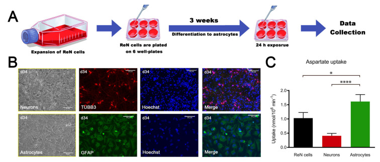
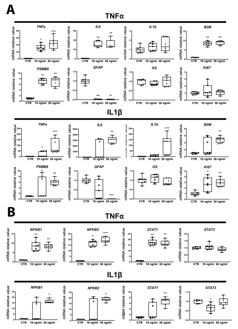
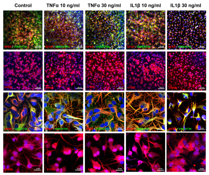
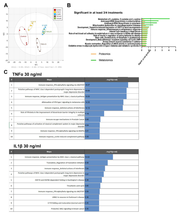
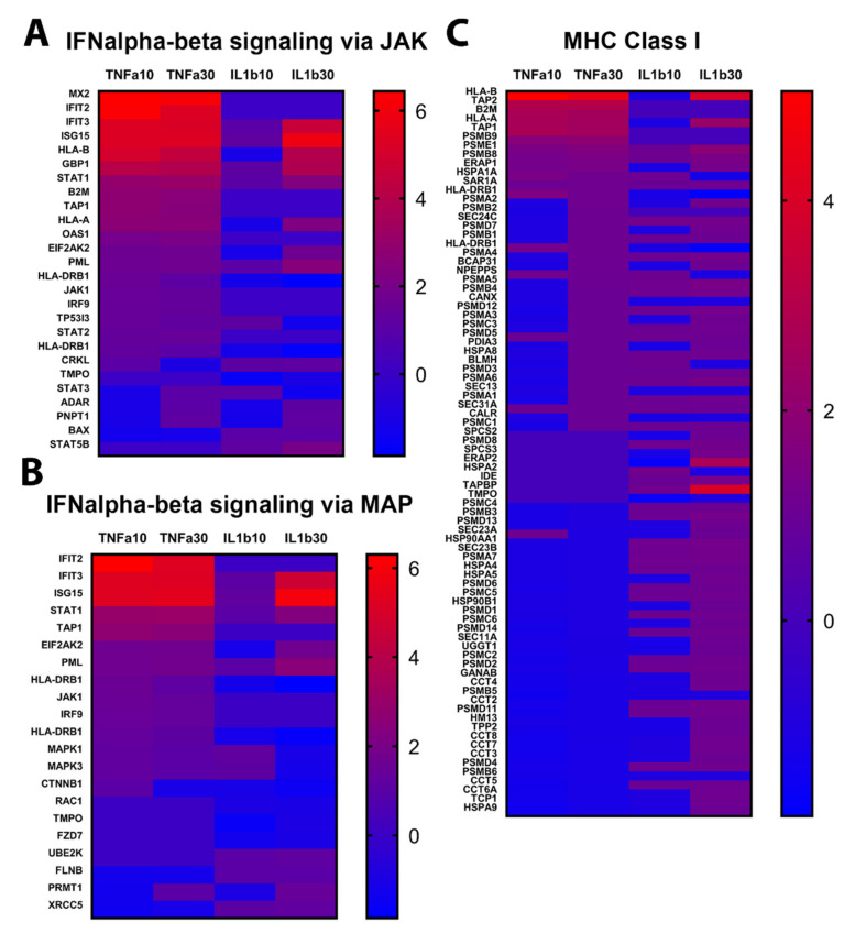
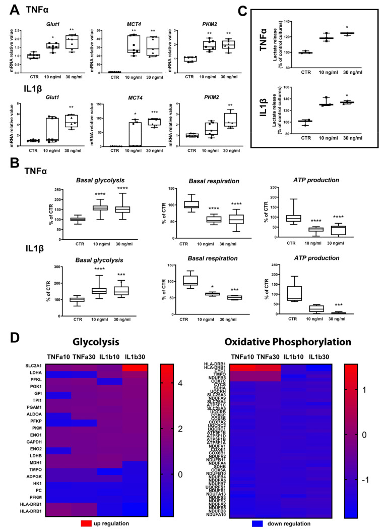
Astrogliosis has been abundantly studied in rodents but relatively poorly in human cells due to limited access to the brain. Astrocytes play important roles in cerebral energy metabolism, and are also key players in neuroinflammation. Astroglial metabolic and inflammatory changes as a function of age have been reported, leading to the hypothesis that mitochondrial metabolism and inflammatory responses are interconnected in supporting a functional switch of astrocytes from neurotrophic to neurotoxic. This study aimed to explore the metabolic changes occurring in astrocytes during their activation. Astrocytes were derived from human ReN cell neural progenitors and characterized. They were activated by exposure to tumor necrosis factor alpha (TNFα) or interleukin 1β (IL1β) for 24 h. Astrocyte reaction and associated energy metabolic changes were assessed by immunostaining, gene expression, proteomics, metabolomics and extracellular flux analyses. ReN-derived astrocytes reactivity was observed by the modifications of genes and proteins linked to inflammation (cytokines, nuclear factor-kappa B (NFκB), signal transducers and activators of transcription (STATs)) and immune pathways (major histocompatibility complex (MHC) class I). Increased NFκB1, NFκB2 and STAT1 expression, together with decreased STAT3 expression, suggest an activation towards the detrimental pathway. Strong modifications of astrocyte cytoskeleton were observed, including a glial fibrillary acidic protein (GFAP) decrease. Astrogliosis was accompanied by changes in energy metabolism characterized by increased glycolysis and lactate release. Increased glycolysis is reported for the first time during human astrocyte activation. Astrocyte activation is strongly tied to energy metabolism, and a possible association between NFκB signaling and/or MHC class I pathway and glycolysis is suggested.

Keywords: astrocytes; astrogliosis; energy metabolism; neuroenergetic; neuroinflammation; reactive astrocytes.


Conflict of interest statement

The authors declare no conflict of interest. The funders had no role in the design of the study; in the collection, analyses, or interpretation of data; in the writing of the manuscript, or in the decision to publish the results.

Figures

Figure 1
Figure 1
ReN cell-derived astrocytes are functional. (A) Scheme of the experiment. (B) Bright field pictures from neurons and astrocytes and immunostaining for TUBB3 (red) and glial fibrillary acidic protein (GFAP) (green); nuclei are stained with Hoechst (blue). (C) Aspartate uptake of ReN cells, neurons and astrocytes. Results are expressed as mean ± SD, n = 10 for ReN cells, n = 9 for neurons, n = 14 for astrocytes, obtained in 3 independent experiments. Kruskal–Wallis test was followed by Dunn’s multiple comparisons test. * p < 0.05, **** p < 0.0001.
Figure 2
Figure 2
Exposure to cytokines induces human ReN-derived astrocyte reactivity. (A) Relative mRNA levels of genes involved in astrocyte reactivity after 24 h of exposure to TNFα or to IL1β. (B) Gene expression regulation of transcription factors after 24 h of exposure to TNFα or to IL1β. Results are displayed as boxplots with data points, for each group n = 7 samples obtained in 2 independent experiments. The line in the box indicates the median, whereas top and bottom of the box represent the 75th and 25th percentiles; whiskers extend from minimum to maximum values. Statistical analysis was performed by using Kruskal–Wallis test followed by Dunn’s multiple comparisons test. * p < 0.05; ** p < 0.01; *** p < 0.001.
Figure 3
Figure 3
Exposure to cytokines induces morphological modifications in human ReN-derived astrocytes. Double immunostaining for GFAP and vimentin and immunostaining for S100B in control and after 24 h of exposure to TNFα or IL1β. First and second rows: low magnification; third and fourth rows: high magnification.
Figure 4
Figure 4
Protein and metabolite pathway analysis after exposure to cytokines. (A) PCA of the metabolites measured by three LC–MS approaches, changing after exposure to TNFα or IL1β (10 and 30 ng/mL) for 24 h. (B) Main pathways enriched in at least 3 out of 4 treatments after pathway analysis with MetaCoreTM v.6.26 (green: metabolomics, orange: proteomics). (C) Top regulated protein pathways found after 24 h of exposure to TNFα (30 ng/mL) or IL1β (30 ng/mL). Proteins and metabolites with fold change >1.2 or <−1.2, and a ratio p-value < 0.05 were kept for pathway enrichment analysis.
Figure 5
Figure 5
Protein enrichment of inflammatory pathways. Abundance of proteins significantly changed after 24 h of exposure to TNFα or IL1β (10 and 30 ng/mL), as compared to control cultures. (A) IFN signaling via JAK pathway. (B) IFN signaling via MAP pathway. (C) MHC class I pathway. Red: upregulated, blue: downregulated. Scales indicate fold change compared to control cultures.
Figure 6
Figure 6
Exposure to cytokines leads to increased glycolysis in ReN-derived astrocytes. (A) Changes in genes related to energy metabolism after 24 h of exposure to TNFα or to IL1β. (B) Basal glycolysis rate after TNFα (n = 48 per group) or IL1β (n = 42 per group); basal respiration after TNFα (control: n = 18, 10 ng/mL: n = 14, 30 ng/mL: n = 13) or IL1β (control: n = 19, 10 ng/mL: n = 5, 30 ng/mL: n = 6); and ATP production after TNFα (control: n = 17, 10 ng/mL: n = 14, 30 ng/mL: n = 13); and after IL1β (control: n = 8, 10 ng/mL: n = 5, 30 ng/mL: n = 6); (C) Lactate release after 24 h of exposure to TNFα or IL1β. n = 3 per group. Results are displayed as boxplots with data points. The line in the box indicates the median, whereas top and bottom of the box represent the 75th and 25th percentiles; whiskers extend from minimum to maximum values. Statistical analysis was performed by using Kruskal–Wallis test followed by Dunn’s multiple comparisons test. * p < 0.05; ** p < 0.01; *** p < 0.001; **** p < 0.0001. (D) Abundance of proteins significantly changed after 24 h of exposure to TNFα or IL1β as compared to control cultures in glycolysis and oxidative phosphorylation pathways. Red: upregulated, blue: downregulated. Scales indicate fold change compared to control cultures.

References

    1. Magistretti P.J., Allaman I. A cellular perspective on brain energy metabolism and functional imaging. Neuron. 2015;86:883–901. doi: 10.1016/j.neuron.2015.03.035. - DOI - PubMed
    1. Magistretti P.J., Pellerin L. Cellular mechanisms of brain energy metabolism and their relevance to functional brain imaging. Philos. Trans. R. Soc. Lond. B Biol. Sci. 1999;354:1155–1163. doi: 10.1098/rstb.1999.0471. - DOI - PMC - PubMed
    1. Baltan S. Can lactate serve as an energy substrate for axons in good times and in bad, in sickness and in health? Metab. Brain Dis. 2015;30:25–30. doi: 10.1007/s11011-014-9595-3. - DOI - PMC - PubMed
    1. Machler P., Wyss M.T., Elsayed M., Stobart J., Gutierrez R., von Faber-Castell A., Kaelin V., Zuend M., San Martin A., Romero-Gomez I., et al. In Vivo Evidence for a Lactate Gradient from Astrocytes to Neurons. Cell Metab. 2016;23:94–102. doi: 10.1016/j.cmet.2015.10.010. - DOI - PubMed
    1. Magistretti P.J., Allaman I. Lactate in the brain: From metabolic end-product to signalling molecule. Nat. Rev. Neurosci. 2018;19:235–249. doi: 10.1038/nrn.2018.19. - DOI - PubMed

MeSH terms

LinkOut - more resources