. 2021 Apr 10;10(4):864.
doi: 10.3390/cells10040864.

Hair-Follicle-Associated Pluripotent (HAP) Stem Cells Can Extensively Differentiate to Tyrosine-Hydroxylase-Expressing Dopamine-Secreting Neurons

Affiliations

Hair-Follicle-Associated Pluripotent (HAP) Stem Cells Can Extensively Differentiate to Tyrosine-Hydroxylase-Expressing Dopamine-Secreting Neurons

Michiko Yamane et al. Cells. .

Abstract

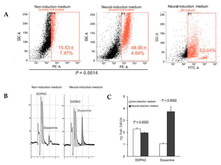
Hair-follicle-associated pluripotent (HAP) stem cells are located in the bulge area of hair follicles from mice and humans and have been shown to differentiate to neurons, glia, keratinocytes, smooth muscle cells, melanocytes and beating cardiac muscle cells in vitro. Subsequently, we demonstrated that HAP stem cells could effect nerve and spinal-cord regeneration in mouse models, differentiating to Schwann cells and neurons in this process. HAP stem cells can be banked by cryopreservation and preserve their ability to differentiate. In the present study, we demonstrated that mouse HAP stem cells cultured in neural-induction medium can extensively differentiate to dopaminergic neurons, which express tyrosine hydroxylase and secrete dopamine. These results indicate that the dopaminergic neurons differentiated from HAP stem cells may be useful in the future to improve the symptoms of Parkinson's disease in the clinic.

Keywords: differentiation; dopamine; hair follicle; hair follicle stem cell area; neural stem cells; neurogenesis; neuron; stem cell.


Conflict of interest statement

The authors declare no conflict of interest.

Figures

Figure 1
Figure 1
Scheme for differentiation of dopaminergic neurons from HAP stem cells. HAP stem cells were initially cultured in neural-induction medium, which was changed as follows: Day 7, neural-progenitor-cells-induction medium (NIM); Day 12, dopaminergic-neuron-differentiation medium (DDM); Day 26, dopaminergic-neuron-maturation medium-1 (DMM1); Day 31, dopaminergic- neuron-maturation medium-2 (DMM2).
Figure 2
Figure 2
HAP stem cells differentiated to dopaminergic neurons. The left panels show HAP stem cells cultured in non-induction medium. The right panels show HAP stem cells cultured in neural-induction medium. (A) Immunofluorescence staining shows that HAP stem cells cultured in neural-induction medium differentiated to βIII-tubulin-positive (red fluorescence) and tyrosine-hydroxylase-positive (green fluorescence) neurons. (blue fluorescence = DAPI). Tyrosine-hydrox-ylase was expressed much more strongly in neural-induction medium. (B) HAP stem cells cultured in neural-induction medium differentiated to dopamine-positive (red fluorescence) dopaminergic neurons. Dopamine was produced much more in neural-induction medium. (C) HAP stem cells cultured in neural-induction medium differentiated to DAT positive (red fluorescence). (D) HAP stem cells cultured in neural-induction medium differentiated to Nurr1 positive (red fluorescence). Scale bar = 100 μm.
Figure 3
Figure 3
Proliferation of dopaminergic neurons differentiated from HAP stem cells. (A) FACS analysis showed that the dopaminergic neurons differentiated efficiently in neural-induction medium. (PE-A = tyrosine-hydroxylase-phycoerythrin. FATC-A = βIII-tubulin). Dopaminergic neurons differentiated from HAP stem cells secreted high levels of dopamine when cultured in neural-induction medium. (B) HPLC shows that the dopaminergic neurons secreted large amounts of dopamine. (C) Dopamine secretion significantly increased in neural-induction medium compared to non-induction medium. DOPAC = 3,4-dihydroxyphenylacetic acid.
Figure 4
Figure 4
Effect of ATP on Ca2+ levels in dopaminergic neurons differentiated from HAP stem cells. (A) ATP caused Ca2+ concentration changes in the dopaminergic neurons with time observed. (B) The line-plots in the bottom panels show the relative fluorescence ratio (F/F0), where F0 is fluorescence before treatment with ATP, and F is fluorescence after treatment. Scale bar = 20 μm.

Similar articles

Cited by

References

    1. Yashiro M., Mii S., Aki R., Hamada Y., Arakawa N., Kawahara K., Hoffman R.M., Amoh Y. From hair to heart: Nestin-expressing hair-follicle-associated pluripotent (HAP) stem cells differentiate to beating cardiac muscle cells. Cell Cycle. 2015;14:2362–2366. doi: 10.1080/15384101.2015.1042633. - DOI - PMC - PubMed
    1. Yamazaki A., Yashiro M., Mii S., Aki R., Hamada Y., Arakawa N., Kawahara K., Hoffman R.M., Amoh Y. Isoproterenol directs hair follicle-associated pluripotent (HAP) stem cells to differentiate in vitro to cardiac muscle cells which can be induced to form beating heart-muscle tissue sheets. Cell Cycle. 2016;15:760–765. doi: 10.1080/15384101.2016.1146837. - DOI - PMC - PubMed
    1. Hoffman R.M. The pluripotency of hair follicle stem cells. Cell Cycle. 2006;5:232–233. doi: 10.4161/cc.5.3.2397. - DOI - PubMed
    1. Amoh Y., Kanoh M., Niiyama S., Kawahara K., Satoh Y., Katsuoka K., Hoffman R.M. Human and mouse hair follicles contain both multipotent and monopotent stem cells. Cell Cycle. 2009;8:176–177. doi: 10.4161/cc.8.1.7342. - DOI - PubMed
    1. Obara K., Tohgi N., Mii S., Hamada Y., Arakawa N., Aki R., Singh S.R., Hoffman R.M., Amoh Y. Hair-follicle-associated pluripotent stem cells derived from cryopreserved intact human hair follicles sustain multilineage differentiation potential. Sci. Rep. 2019;9:9326. doi: 10.1038/s41598-019-45740-9. - DOI - PMC - PubMed

Publication types

LinkOut - more resources