Review
. 2021 Apr 10;10(4):865.
doi: 10.3390/cells10040865.

Atherothrombosis in Acute Coronary Syndromes-From Mechanistic Insights to Targeted Therapies

Affiliations
Review

Atherothrombosis in Acute Coronary Syndromes-From Mechanistic Insights to Targeted Therapies

Chinmay Khandkar et al. Cells. .

Abstract

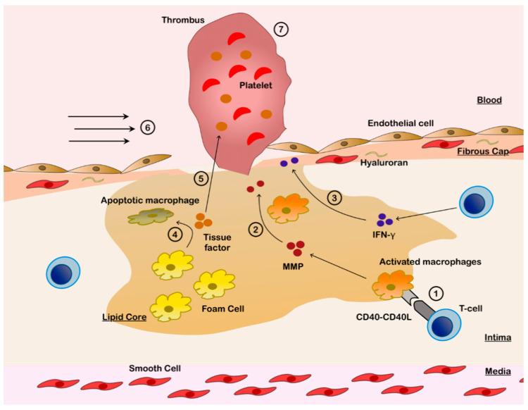
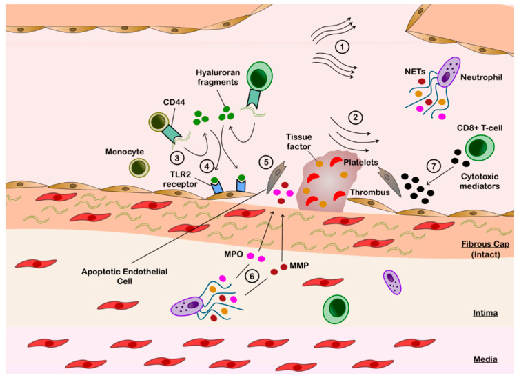
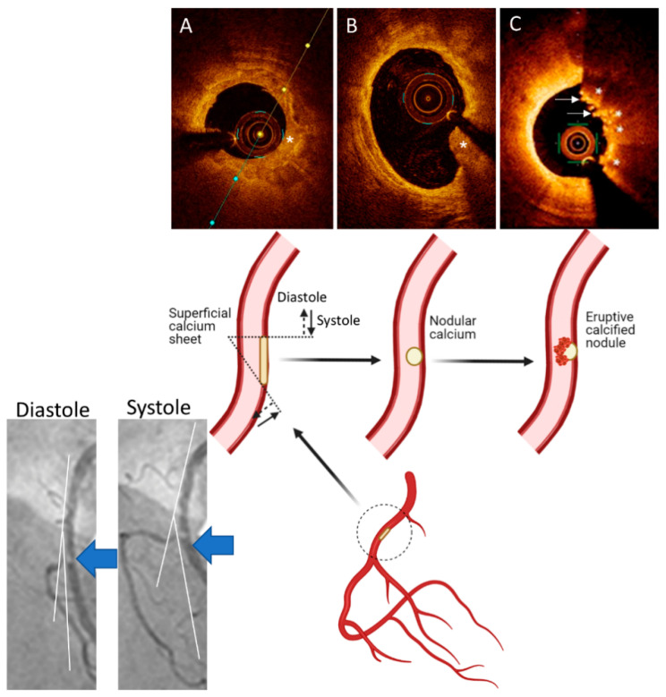
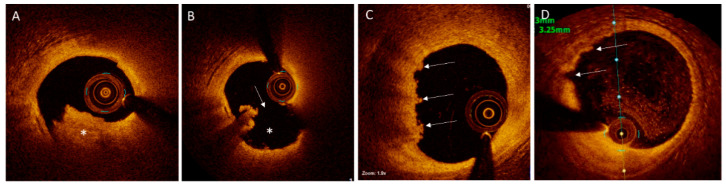
The atherothrombotic substrates for acute coronary syndromes (ACS) consist of plaque ruptures, erosions and calcified nodules, while the non-atherothrombotic etiologies, such as spontaneous coronary artery dissection, coronary artery spasm and coronary embolism are the rarer causes of ACS. The purpose of this comprehensive review is to (1) summarize the histopathologic insights into the atherothrombotic plaque subtypes in acute ACS from postmortem studies; (2) provide a brief overview of atherogenesis, while mainly focusing on the events that lead to plaque destabilization and disruption; (3) summarize mechanistic data from clinical studies that have used intravascular imaging, including high-resolution optical coherence tomography, to assess culprit plaque morphology and its underlying pathobiology, especially the newly described role of innate and adaptive immunity in ACS secondary to plaque erosion; (4) discuss the utility of intravascular imaging for effective treatment of patients presenting with ACS by percutaneous coronary intervention; and (5) discuss the opportunities that these mechanistic and imaging insights may provide for more individualized treatment of patients with ACS.

Keywords: acute coronary syndromes; calcified nodule; optical coherence tomography; percutaneous coronary intervention; plaque erosion; plaque rupture.


Conflict of interest statement

Madhavan is supported by an institutional grant by the National Heart, Lung, and Blood Institute of the US National Institutes of Health to Columbia University Irving Medical Center (T32 HL007854). The other authors declare no conflicts relevant to the content of this manuscript.

Figures

Figure 1
Figure 1
Pathobiology of Plaque Rupture. 1. Infiltrating type 1 T-helper cells activate macrophages in lipid core via CD40L-CD40 interaction. 2. Activated macrophages release matrix metalloproteinases (MMPs), which disrupt the fibrous cap and induce endothelial apoptosis. 3. Infiltrating type 1 T-helper cells release interferon-γ (IFN-γ), which inhibits smooth muscle cells and thereby weakens the fibrous cap. 4. Foam cells become apoptotic due to CD40 stimulation, releasing tissue factor. 5. Tissue factor, which has potent thrombogenic potential, is released into the blood stream. 6. Endothelial apoptosis is augmented by mechanical stress from increased shear forces, especially at the plaque edges, resulting in fibrous cap rupture. 7. Thrombus is formed over the exposed thrombogenic lipidic core.
Figure 2
Figure 2
Pathobiology of Plaque Erosion. 1. Impaired laminar flow secondary to bifurcation point. 2. Low shear forces downstream of plaque disrupt cell junctions to induce endothelial apoptosis and mobilize hyaluronan from the fibrous cap. 3. Exposed hyaluronan binds to the circulating monocytes via CD44 and is then hydrolyzed by hyalurorannidase-2 into hyaluronan fragments. 4. Hyaluronan fragments bind to the toll like receptor 2 (TLR2) on endothelial cells. 5. TLR2-stimulated endothelial cells are induced into apoptosis. 6. Endothelial denudation attracts neutrophils, which release neutrophil extracellular traps (NETs) containing metalloproteinase (MMP) and myeloperoxidase (MPO), to augment endothelial apoptosis. Tissue factor is also released in NETs. 7. Infiltrating CD8+ T cells release granzyme A, granulysin and perforin to further disrupt the endothelial layer. 8. Endothelial disruption exposes the underlying fibrous cap, which results in activation of the external coagulation cascade in conjunction with the thrombogenic tissue factor to form thrombus.
Figure 3
Figure 3
A Working Hypothesis for Formation of Eruptive Calcified Nodules. Superficial calcified sheets (asterisk) (A) located in the arterial segments with hinge movement, e.g., in the mid segment of the right coronary artery (large blue arrows on the angiographic pictures and inset), are subject to cyclic mechanical forces during systole and diastole, which could weaken and fragment the sheets of calcium, thus resulting in protruding nodular calcium (asterisk) (B) that is surrounded by fibrin. These nodules eventually erupt through the plaque surface ((C), asterisks), causing disruption in the intima, with superimposition of thrombus ((C), arrows). Parts of the figure are adapted from Lee et al. [55] with permission.
Figure 4
Figure 4
Plaques with Disrupted or Intact Fibrous Cap on Optical Coherence Tomography. (A). Mixed white and red thrombus (asterisk) in the left anterior descending (LAD) artery of a patient presenting with ST-segment elevation myocardial infarction. (B). After aspiration thrombectomy, a disrupted fibrous cap (arrow) with an empty crater (asterisk), typical for the ruptured plaques, is visualized. (C). “Definite” plaque erosion in the LAD artery of a patient presenting with non-ST-segment elevation myocardial infarction. Irregular plaque surface and predominantly white thrombi (arrows) are noted. (D). “Probable” plaque erosion is detected in a patient presenting non-ST-segment elevation myocardial infarction by the presence of irregular plaque surface (arrows) without overlying thrombus in the LAD artery.

Similar articles

Cited by

References

    1. Burke A.P., Farb A., Malcom G.T., Liang Y.H., Smialek J., Virmani R. Coronary risk factors and plaque morphology in men with coronary disease who died suddenly. N. Engl. J. Med. 1997;336:1276–1282. doi: 10.1056/NEJM199705013361802. - DOI - PubMed
    1. Farb A., Tang A.L., Burke A.P., Sessums L., Liang Y., Virmani R. Sudden coronary death. Frequency of active coronary lesions, inactive coronary lesions, and myocardial infarction. Circulation. 1995;92:1701–1709. doi: 10.1161/01.CIR.92.7.1701. - DOI - PubMed
    1. Virmani R., Frank D.K., Allen P.B., Farb A., Stephen M.S. Lessons From Sudden Coronary Death. Arterioscler. Thromb. Vasc. Biol. 2000;20:1262–1275. doi: 10.1161/01.ATV.20.5.1262. - DOI - PubMed
    1. Bentzon J.F., Otsuka F., Virmani R., Falk E. Mechanisms of Plaque Formation and Rupture. Circ. Res. 2014;114:1852–1866. doi: 10.1161/CIRCRESAHA.114.302721. - DOI - PubMed
    1. Stary H.C., Chandler A.B., Glagov S., Guyton J.R., Insull W., Jr., Rosenfeld M.E., Schaffer S.A., Schwartz C.J., Wagner W.D., Wissler R.W. A definition of initial, fatty streak, and intermediate lesions of atherosclerosis. A report from the Committee on Vascular Lesions of the Council on Arteriosclerosis, American Heart Association. Circulation. 1994;89:2462–2478. doi: 10.1161/01.CIR.89.5.2462. - DOI - PubMed

Publication types

MeSH terms

LinkOut - more resources