Review
. 2021 Apr 17;22(8):4164.
doi: 10.3390/ijms22084164.

A Walk in the Memory, from the First Functional Approach up to Its Regulatory Role of Mitochondrial Bioenergetic Flow in Health and Disease: Focus on the Adenine Nucleotide Translocator

Affiliations
Review

A Walk in the Memory, from the First Functional Approach up to Its Regulatory Role of Mitochondrial Bioenergetic Flow in Health and Disease: Focus on the Adenine Nucleotide Translocator

Anna Atlante et al. Int J Mol Sci. .

Abstract

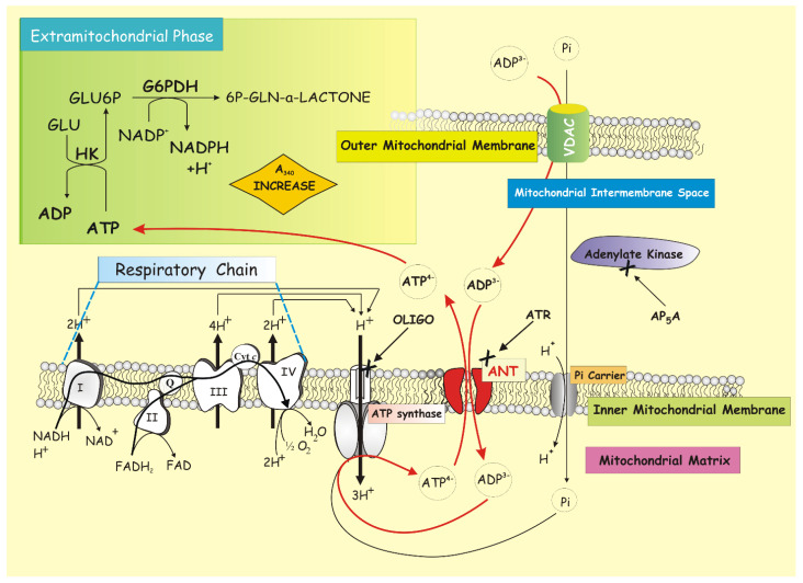
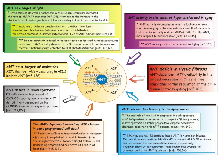
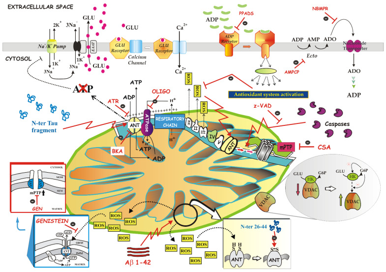
The mitochondrial adenine nucleotide translocator (ANT) plays the fundamental role of gatekeeper of cellular energy flow, carrying out the reversible exchange of ADP for ATP across the inner mitochondrial membrane. ADP enters the mitochondria where, through the oxidative phosphorylation process, it is the substrate of Fo-F1 ATP synthase, producing ATP that is dispatched from the mitochondrion to the cytoplasm of the host cell, where it can be used as energy currency for the metabolic needs of the cell that require energy. Long ago, we performed a method that allowed us to monitor the activity of ANT by continuously detecting the ATP gradually produced inside the mitochondria and exported in the extramitochondrial phase in exchange with externally added ADP, under conditions quite close to a physiological state, i.e., when oxidative phosphorylation takes place. More than 30 years after the development of the method, here we aim to put the spotlight on it and to emphasize its versatile applicability in the most varied pathophysiological conditions, reviewing all the studies, in which we were able to observe what really happened in the cell thanks to the use of the "ATP detecting system" allowing the functional activity of the ANT-mediated ADP/ATP exchange to be measured.

Keywords: ATP detecting system; adenine nucleotide translocator; disease; mitochondria; physiological role; transport.


Conflict of interest statement

The authors declare no conflict of interest.

Figures

Figure 1
Figure 1
Functional activity of ANT-mediated ADP/ATP exchange revealed by an ATP detecting system. The ATP detecting system (ATP D.S.), depicted in the upper left panel, reveals the appearance of ATP in the extramitochondrial phase following the addition of ADP to the mitochondria and the synthesis of ATP via OXPHOS. As shown in the picture, since the activity of the ANT-dependent electrogenic ADP/ATP exchange strictly depends on the mitochondrial membrane potential, mitochondria must be actively “respiring and phosphorylating,” i.e., the activity of mRC that generates the electrochemical gradient transmembrane, and ATP synthesis must be closely coupled for the exchange to occur.
Figure 2
Figure 2
ANT is the target of light or molecules or even of the two Alzheimer’s proteins and its activity is impaired under various pathological conditions affecting the host cell.
Figure 3
Figure 3
The picture collects data from several studies on the role of ANT and its functionality in the neurons sentenced to death for apoptosis or necrosis. In particular, the dual role of ANT, harboring two opposite functions, i.e., a vital function important to ensuring the life of the cell, and the other lethal—such as protein involved in the participation of mPTP opening—leading the cell to death, is depicted. The effect of NH2htau and Aβ1-42 peptides impairing mitochondrial ANT-1 in Alzheimer’s disease is also shown. The data represented here refer to the reference numbers [145,146,147,148,149,150,151,152,153].

Similar articles

Cited by

References

    1. Palade G.E. An electron microscope study of the mitochondrial structure. J. Histochem. Cytochem. 1953;1:188–211. doi: 10.1177/1.4.188. - DOI - PubMed
    1. Vogel F., Bornhovd C., Neupert W., Reichert A.S. Dynamic subcompartmentalization of the mitochondrial inner membrane. J. Cell Biol. 2006;175:237–247. doi: 10.1083/jcb.200605138. - DOI - PMC - PubMed
    1. Perkins G., Renken C., Martone M.E., Young S.J., Ellisman M., Frey T. Electron tomography of neuronal mitochondria: Three-dimensional structure and organization of cristae and membrane contacts. J. Struct. Biol. 1997;119:260–272. doi: 10.1006/jsbi.1997.3885. - DOI - PubMed
    1. Appelhans T., Busch K.B. Dynamic imaging of mitochondrial membrane proteins in specific sub-organelle membrane locations. Biophys. Rev. 2017;9:345–352. doi: 10.1007/s12551-017-0287-1. - DOI - PMC - PubMed
    1. Chacinska A., Koehler C.M., Milenkovic D., Lithgow T., Pfanner N. Importing mitochondrial proteins: Machineries and mechanisms. Cell. 2009;138:628–644. doi: 10.1016/j.cell.2009.08.005. - DOI - PMC - PubMed

MeSH terms

Substances

LinkOut - more resources