Review
. 2021 Apr 17;12(4):591.
doi: 10.3390/genes12040591.

Structures of the ß-Keratin Filaments and Keratin Intermediate Filaments in the Epidermal Appendages of Birds and Reptiles (Sauropsids)

Affiliations
Review

Structures of the ß-Keratin Filaments and Keratin Intermediate Filaments in the Epidermal Appendages of Birds and Reptiles (Sauropsids)

David A D Parry. Genes (Basel). .

Abstract

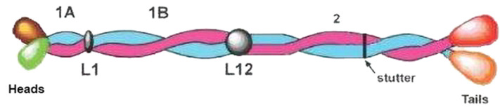
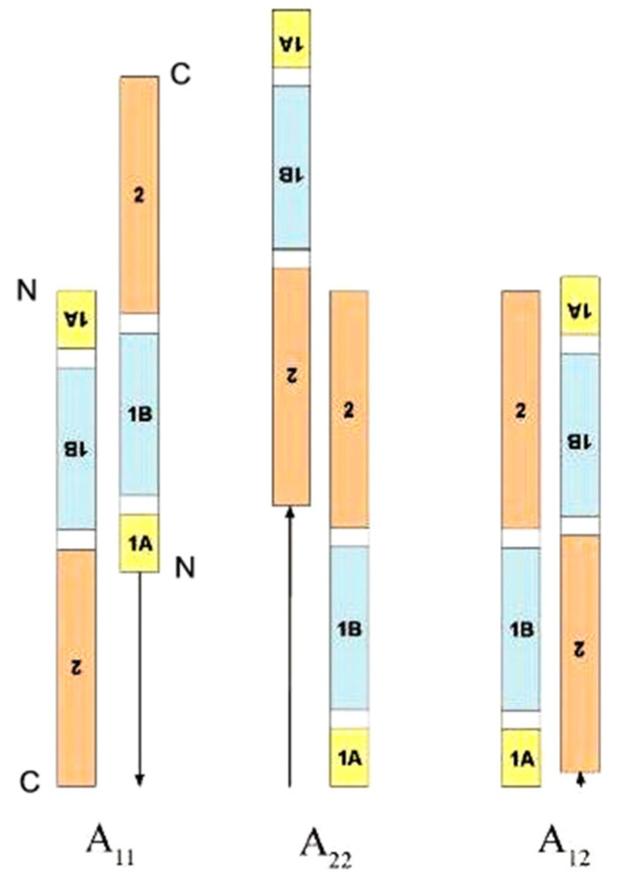
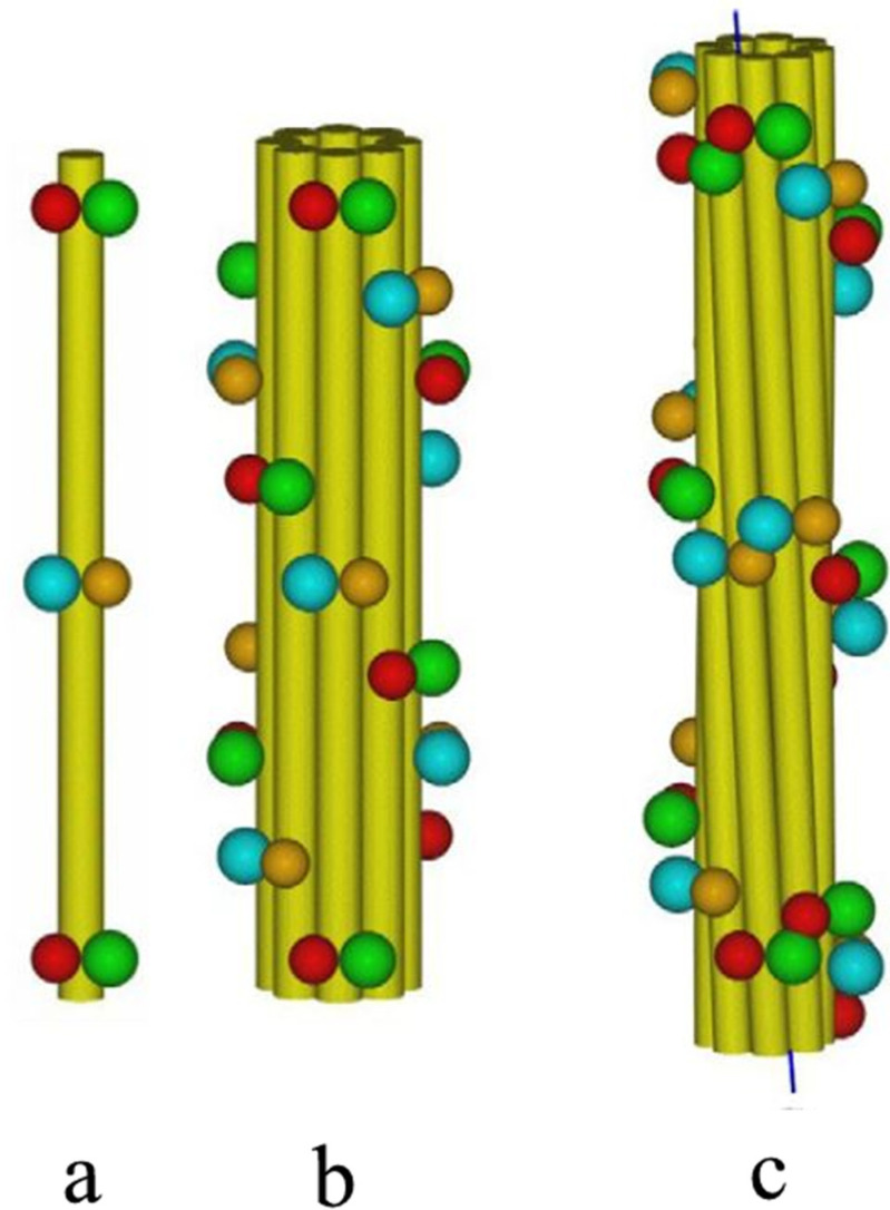
The epidermal appendages of birds and reptiles (the sauropsids) include claws, scales, and feathers. Each has specialized physical properties that facilitate movement, thermal insulation, defence mechanisms, and/or the catching of prey. The mechanical attributes of each of these appendages originate from its fibril-matrix texture, where the two filamentous structures present, i.e., the corneous ß-proteins (CBP or ß-keratins) that form 3.4 nm diameter filaments and the α-fibrous molecules that form the 7-10 nm diameter keratin intermediate filaments (KIF), provide much of the required tensile properties. The matrix, which is composed of the terminal domains of the KIF molecules and the proteins of the epidermal differentiation complex (EDC) (and which include the terminal domains of the CBP), provides the appendages, with their ability to resist compression and torsion. Only by knowing the detailed structures of the individual components and the manner in which they interact with one another will a full understanding be gained of the physical properties of the tissues as a whole. Towards that end, newly-derived aspects of the detailed conformations of the two filamentous structures will be discussed and then placed in the context of former knowledge.

Keywords: X-ray fiber diffraction; corneous ß-proteins; keratin intermediate filaments.


Conflict of interest statement

The author declares that he has no known competing financial interests or personal relationships that could have appeared to influence the work reported in this paper.

Figures

Figure 1
Figure 1
Phylogenetic classification of the sauropsids. The branching times are measured in millions of years (Mya) and are based on studies of mitochondrial DNA [1]. Figure reproduced from [5] with permission of Elsevier.
Figure 2
Figure 2
Transmission electron micrograph of a cross-section of feather keratin showing filaments about 3–4 nm in diameter [6]. Figure reproduced from [31] with permission of Elsevier.
Figure 3
Figure 3
High angle X-ray diffractions patterns of (a) seagull feather rachis (Larus novae-hollandiae), which shows features of a ß-conformation, and (b) porcupine quill (Hystrix cristata), which is based on the α-conformation with its characteristic 0.51 nm meridional reflection and the 0.98 nm equatorial and near-equatorial reflections, (c,d) show the same seagull feather rachis before and after pressing in steam to remove the lateral organisation between the filaments and (e) goanna claw (Varanus varius). (b) reproduced from [43] with permission of Elsevier; (ae) reproduced from [44] with permission of Elsevier.
Figure 4
Figure 4
Sequence comparisons of the CBPs from the sauropsids reveal that the N-terminal domain is comprised of subdomain A in the archelosaurs, but subdomains A and B in the squamates and rhynchocephalia. The C-terminal domain, however, comprises subdomains C and D in all of the sauropsids. The sequence characteristics of each subdomain are listed in the text. The approximate size ranges of the subdomains are listed. The central 34-residue domain adopts a twisted ß-sheet structure with three inner strands and two partial outer ones that assembles in an antiparallel manner with a similar sheet from a different chain to form a ß-sandwich. In turn, these assemble axially with a four-fold screw symmetry to form a filamentous structure of diameter 3.4 nm. Figure reproduced from [58] with permission of Springer.
Figure 5
Figure 5
Schematic diagram of the ß-sandwich formed from the conserved 34-residue stretch of sequence in each of two CBP chains. Each sandwich is comprised of a pair of right-handed twisted antiparallel ß-sheets (one is drawn blue and the other is yellow) and these are related to one another by a perpendicular dyad axis of rotation (marked alternatively by red circles and red lines). Axial assembly of the ß-sandwiches through the application of a left-handed four-fold screw axis generates a 3.4 nm diameter filament of pitch length 9.6 nm and axial rise 2.4 nm. Figure reproduced from [58] with permission of Springer.
Figure 6
Figure 6
Amino acid sequence of the tuatara chain with four 34-residue repeats. Each repeat is marked with a solid black line. Bars at either end (turquoise) indicate short conserved regions believed to have a role in stabilization or assembly. Color coding for selected amino acids is as follows: cysteine (yellow), proline (magenta), glycine (grey), tyrosine (green), acidic residues (red), and basic residues (blue). Figure reproduced from [5] with permission of Elsevier.
Figure 7
Figure 7
The structure proposed for the chains with four 34-residue repeats contains two ß-sandwiches (pink/green and blue/orange). One occurs in each of two neighboring filaments and are linked by the sequence connecting repeats 2 and 3 (represented by a wavy line). The linker is believed to adopt a ß-like sheet characterized by stretches of sequence with a serine-X repeat. Figure reproduced from [67] with permission of Elsevier.
Figure 8
Figure 8
An axial projection of neighboring filaments in feather keratin showing their common orientation, as deduced from the row line pattern seen in the X-ray diffraction pattern. The ß-sandwiches are inclined at an angle of 17° to the plane of the sheet and this results in a ß-sheet in one filament lining up with a ß-sheet in a neighboring filament. Reproduced from [63] with permission of Elsevier.
Figure 9
Figure 9
Electron micrograph of a cross-section of fine Merino wool showing the quasi-hexagonal packing of keratin intermediate filaments about 7–10 nm in diameter embedded in an osmiophilic matrix [70]. The magnification bar is 100 nm. The inset (at the same magnification) shows porcupine quill post-stained with potassium permanganate. The KIF have a ring structure. Figure reproduced from [71] with permission of Springer.
Figure 10
Figure 10
Schematic structure of the KIF molecule with a Type I and a Type II chain in axial register and parallel to one another. The 1A, 1B, and 2 segments have a heptad substructure and form a left-handed two-stranded coiled-coil with connecting linkers L1 and L12. At the N-terminal end of segment 2 the constituent chains have a hendecad repeat which causes the chains to lie approximately parallel to the axis rather than being coiled about it. The N-terminal (head) and C-terminal (tail) domains enclose the central rod domain. Figure reproduced from [71] with permission of Springer.
Figure 11
Figure 11
In total, three modes of molecular assembly are found in IF [82,87]. These are termed A11, A22, and A12 and involve the approximate axial alignment of antiparallel 1B segments, antiparallel 2 segments and antiparallel rod domains, respectively. The axial staggers for each mode are measured from the N-terminus of segment 1A in an up-pointing molecule. Figure reproduced from [71] with permission of Elsevier.
Figure 12
Figure 12
Models of (a) a single protofilament in the reduced state, (b) the reduced IF with eight protofilaments arranged on a ring of radius 3.5 nm, and (c) the oxidized IF, again with eight protofilaments, arranged on a ring of radius 3.0 nm. The yellow cylinders represent the rod domain of the IF molecules, whereas the spheres represent the terminal domains: green = both heads of the Up strands, red = both tails of the Up strands, blue = both heads of the Down strands, and orange = both tails of the Down strands. In (b) the terminal domains in the reduced structure are arranged on a two-start left-handed helix, thereby giving rise to a diagonal banding with a spacing of 22 nm. In contrast, in (c) the terminal domains in the oxidized structure with its dislocated lattice lie on a one-start helix of pitch length 23.5 nm. Figure reproduced from [97] with permission of Springer.

Similar articles

Cited by

References

    1. Rest J.S., Ast J.C., Austin C.C., Wadell P.J., Tibbetts E.A., Hay J.M., Mindell D.P. Molecular systematics of primary reptilian lineages and the tuatara mitochondrial genome. Mol. Phylogenet. Evol. 2003;29:289–297. doi: 10.1016/S1055-7903(03)00108-8. - DOI - PubMed
    1. Rice R.H., Winters B.R., Durbin-Johnson B.P., Rocke D.M. Chicken corneocyte cross-linked proteome. J. Proteome Res. 2013;12:771–776. doi: 10.1021/pr301036k. - DOI - PMC - PubMed
    1. Strasser B., Mlitz V., Hermann M., Tschachler E., Eckhart L. Convergent evolution of cysteine-rich proteins in feathers and hair. BMC Evol. Biol. 2015;15:82. doi: 10.1186/s12862-015-0360-y. - DOI - PMC - PubMed
    1. Wu P., Ng C.S., Yan J., Lai Y.C., Chen C.K., Lai Y.T., Wu S.M., Chen J.J., Luo W., Widelitz R.B., et al. Topographical mapping of α- and β-keratins on developing chicken skin integument: Functional interaction and evolutionary perspectives. Proc. Natl. Acad. Sci. USA. 2015;122:E6770–E6779. doi: 10.1073/pnas.1520566112. - DOI - PMC - PubMed
    1. Parry D.A.D., Fraser R.D.B., Alibardi L., Rutherford K., Gemmell N. Molecular structure of sauropsid β-keratins from tuatara (Sphenodon punctatus) J. Struct. Biol. 2019;207:21–28. doi: 10.1016/j.jsb.2019.04.008. - DOI - PubMed

LinkOut - more resources