Review
. 2021 Apr 17;22(8):4170.
doi: 10.3390/ijms22084170.

The Neutrophil Secretome as a Crucial Link between Inflammation and Thrombosis

Affiliations
Review

The Neutrophil Secretome as a Crucial Link between Inflammation and Thrombosis

María Amparo Blanch-Ruiz et al. Int J Mol Sci. .

Abstract

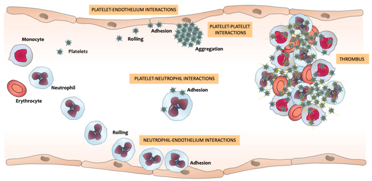
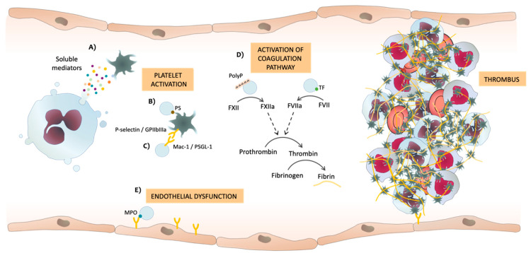
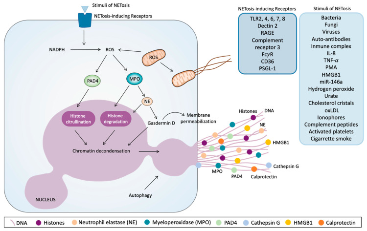
Cardiovascular diseases are a leading cause of death. Blood-cell interactions and endothelial dysfunction are fundamental in thrombus formation, and so further knowledge of the pathways involved in such cellular crosstalk could lead to new therapeutical approaches. Neutrophils are secretory cells that release well-known soluble inflammatory signaling mediators and other complex cellular structures whose role is not fully understood. Studies have reported that neutrophil extracellular vesicles (EVs) and neutrophil extracellular traps (NETs) contribute to thrombosis. The objective of this review is to study the role of EVs and NETs as key factors in the transition from inflammation to thrombosis. The neutrophil secretome can promote thrombosis due to the presence of different factors in the EVs bilayer that can trigger blood clotting, and to the release of soluble mediators that induce platelet activation or aggregation. On the other hand, one of the main pathways by which NETs induce thrombosis is through the creation of a scaffold to which platelets and other blood cells adhere. In this context, platelet activation has been associated with the induction of NETs release. Hence, the structure and composition of EVs and NETs, as well as the feedback mechanism between the two processes that causes pathological thrombus formation, require exhaustive analysis to clarify their role in thrombosis.

Keywords: extracellular vesicles; inflammation; neutrophil; neutrophil extracellular traps; platelets; secretome; thrombosis.


Conflict of interest statement

None of the authors report any potential conflict of interest. The funders had no role in the design of the study; in the collection, analyses, or interpretation of data; in the writing of the manuscript, or in the decision to publish the results.

Figures

Figure 1
Figure 1
Neutrophils’, platelets’, and endothelial cells’ interactions that promote the transition from inflammation to thrombosis. Upon an inflammatory signal, neutrophils circulating across the vessels reduce their flow velocity and start rolling along endothelial cells, eventually adhering to them. This process can induce the activation and recruitment of further neutrophils, in addition to endothelium activation or dysfunction, which can trigger platelet adhesion to the endothelium and to neutrophils. The interactions between these three types of vascular cells, and with circulating monocytes and erythrocytes, trigger the thrombus formation that can produce vessel occlusion.
Figure 2
Figure 2
Potential mechanisms by which soluble mediators or neutrophil extracellular vesicles (EVs) released by neutrophils can induce thrombosis. (A) Soluble mediators released by neutrophils, (B) phosphatidylserine (PS) and (C) different adhesion molecules present in the membrane of EVs interact with platelets, thereby inducing platelet activation and aggregation. (D) EVs activate both intrinsic and extrinsic coagulation pathways. Polyphosphates (PolyP) and tissue factor (TF) in EVs can activate the coagulation cascade, thus producing thrombin from its inactive form of pro-thrombin, and thrombin finally induces the formation of fibrin from fibrinogen. (E) Myeloperoxidase (MPO) in EVs can induce endothelial dysfunction, which triggers platelet activation and aggregation. F: factor; GP: glycoprotein; PSGL-1: P-selectin glycoprotein ligand-1.
Figure 3
Figure 3
Molecular pathways of the release of neutrophil extracellular traps (NETs). Different stimuli and several receptors can induce NETs’ release. One of the first intracellular events that initiate this phenomenon is reactive oxygen species (ROS) production via nicotinamide adenine dinucleotide phosphate (NADPH) or by mitochondria. ROS induce the activation and translocation to the nucleus of peptidyl arginine deiminase 4 (PAD4), which citrullinates histones and contributes to chromatin decondensation. On the other hand, ROS induce myeloperoxidase (MPO) activation, thus promoting neutrophil elastase (NE) release from granules and their translocation to the nucleus, where NE can degrade histones and promotes chromatin decondensation. NE also activates proteolytically gasdermin D, which can facilitate NE translocation to the nucleus, thus increasing chromatin decondensation. Gasdermin D promotes the disassembly of the nuclear envelope, after which nuclear chromatin decondenses into the cytoplasm, mixing with the cytoplasmatic and granule components and also the permeabilization of plasma membrane, thus allowing NETs to expand into the extracellular space. Autophagy is another cellular mechanism that can trigger the release of NETs. DNA: Deoxyribonucleic acid. TLR: Toll-like receptor. RAGE: Receptor for advanced glycation end products. CD: cluster of differentiation. PSGL-1: P-selectin glycoprotein ligand-1. IL: interleukin. TNF-α: Tumor necrosis factor alpha. PMA: Phorbol 12-myristate 13-acetate. HMGB1: high-mobility group protein B1. miR-146a: micro Ribonucleic acid 146a. oxLDL: oxidized low-density lipoprotein.

References

    1. Şahin B., İlgün G. Risk Factors of Deaths Related to Cardiovascular Diseases in World Health Organization (WHO) Member Countries. Health Soc. Care Commun. 2020 doi: 10.1111/hsc.13156. - DOI - PubMed
    1. Roth G.A., Mensah G.A., Johnson C.O., Addolorato G., Ammirati E., Baddour L.M., Barengo N.C., Beaton A.Z., Benjamin E.J., Benziger C.P., et al. Global Burden of Cardiovascular Diseases and Risk Factors, 1990-2019: Update from the GBD 2019 Study. J. Am. Coll. Cardiol. 2020;76:2982–3021. doi: 10.1016/j.jacc.2020.11.010. - DOI - PMC - PubMed
    1. Ay C., Pabinger I., Cohen A.T. Cancer-Associated Venous Thromboembolism: Burden, Mechanisms, and Management. Thromb. Haemost. 2017;117:219–230. doi: 10.1160/TH16-08-0615. - DOI - PubMed
    1. Honig L.S., Tang M., Albert S., Costa R., Luchsinger J., Manly J., Stern Y., Mayeux R. Stroke and the Risk of Alzheimer Disease. Arch. Neurol. 2003;60:1707–1712. doi: 10.1001/archneur.60.12.1707. - DOI - PubMed
    1. Zenaro E., Pietronigro E., Della Bianca V., Piacentino G., Marongiu L., Budui S., Turano E., Rossi B., Angiari S., Dusi S., et al. Neutrophils Promote Alzheimer’s Disease-Like Pathology and Cognitive Decline Via LFA-1 Integrin. Nat. Med. 2015;21:880–886. doi: 10.1038/nm.3913. - DOI - PubMed

LinkOut - more resources