Review
. 2021 Apr 15;22(8):4067.
doi: 10.3390/ijms22084067.

Pharmacological Modulators of Autophagy as a Potential Strategy for the Treatment of COVID-19

Affiliations
Review

Pharmacological Modulators of Autophagy as a Potential Strategy for the Treatment of COVID-19

Gustavo José da Silva Pereira et al. Int J Mol Sci. .

Abstract

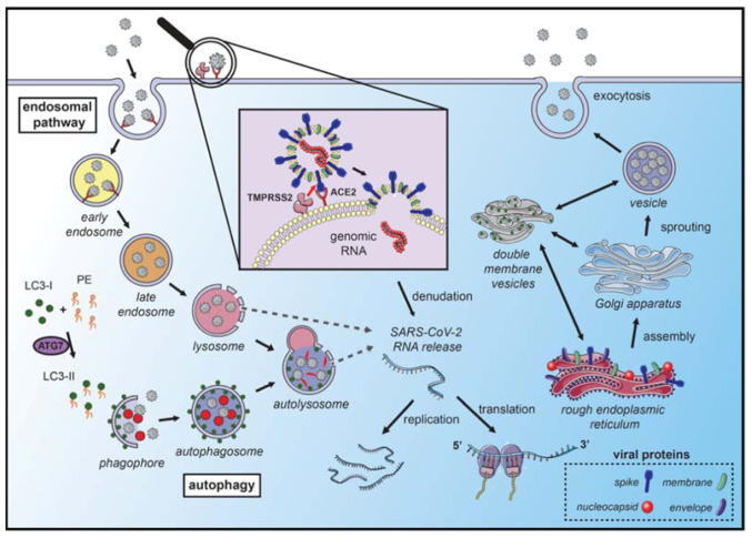
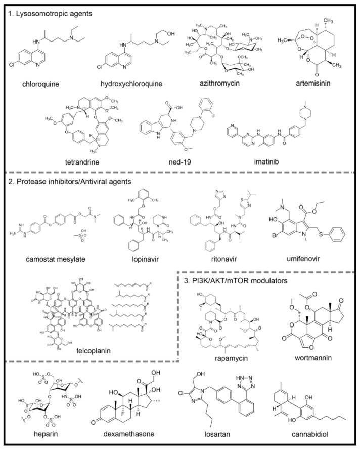
The family of coronaviruses (CoVs) uses the autophagy machinery of host cells to promote their growth and replication; thus, this process stands out as a potential target to combat COVID-19. Considering the different roles of autophagy during viral infection, including SARS-CoV-2 infection, in this review, we discuss several clinically used drugs that have effects at different stages of autophagy. Among them, we mention (1) lysosomotropic agents, which can prevent CoVs infection by alkalinizing the acid pH in the endolysosomal system, such as chloroquine and hydroxychloroquine, azithromycin, artemisinins, two-pore channel modulators and imatinib; (2) protease inhibitors that can inhibit the proteolytic cleavage of the spike CoVs protein, which is necessary for viral entry into host cells, such as camostat mesylate, lopinavir, umifenovir and teicoplanin and (3) modulators of PI3K/AKT/mTOR signaling pathways, such as rapamycin, heparin, glucocorticoids, angiotensin-converting enzyme inhibitors (IECAs) and cannabidiol. Thus, this review aims to highlight and discuss autophagy-related drugs for COVID-19, from in vitro to in vivo studies. We identified specific compounds that may modulate autophagy and exhibit antiviral properties. We hope that research initiatives and efforts will identify novel or "off-label" drugs that can be used to effectively treat patients infected with SARS-CoV-2, reducing the risk of mortality.

Keywords: COVID-19; autophagy; pharmacology.


Conflict of interest statement

The authors declare no conflict of interest.

Figures

Figure 1
Figure 1
Coronavirus hijacks autophagy machinery to promote their replication. SARS-CoVs bind to the angiotensin-converting enzyme 2 (ACE2) receptor on the membrane surface and enter the host cell. The fusion with the membrane and the release of the genomic RNA into the cytoplasm occurs after the cleavage of the spike (S) protein, which can occur in several locations. S protein cleavage occurs on the cell membrane surface by the transmembrane protease serine 2 (TMPRSS2), which is associated with the ACE2 receptor, or by cathepsin-L and cysteine proteases in the endosomal system. The acidic pH in the lysosomes is necessary for the activity of cathepsin-L and S protein cleavage. Next, the endosomal cargo converges with the autophagic vacuoles in the lysosomes. Coronavirus nonstructural proteins colocalize with microtubule-associated proteins 1A/1B light chain 3A (LC3-II) in the endomembrane system, suggesting that autophagy plays a role in amplifying coronavirus replication. After fusion with the membrane, the genomic RNA is released and stripped of the nucleocapsid protein. Viral proteins are translated in the endoplasmic reticulum, which promotes the rearrangement of endoplasmic reticulum membranes and the formation of double-membrane vesicles, which are also localized with LC3 and autophagy-related proteins. The newly synthesized genomic RNA is then assembled into virions in intermediate compartments located between the endoplasmic reticulum and the Golgi apparatus and moves through the secretory pathway of the host and eventually released by exocytosis (the illustration was produced using the smart servier medical art vectors for publications and presentations licensed under the Creative Commons (CC BY 3.0)) [93].
Figure 2
Figure 2
Chemical structures of potential autophagy-related drugs for SARS-CoV-2 infection. The drugs were divided in three groups according to their effects on the autophagy signaling pathway and possible effect against SARS-CoV-2 infection. The lysosomotropic agents (1) can prevent coronavirus infection by alkalinizing the acid pH in the endolysosomal system; some examples are chloroquine, hydroxychloroquine, azithromycin, artemisinin, two-pore channel antagonists (such as tetrandrine and ned-19) and imatinib. The protease inhibitors/antiviral agents (2) can inhibit the proteolytic cleavage of the spike coronavirus protein, which is necessary for viral entry into host cells; some examples are camostat mesylate, lopinavir, ritonavir, umifenovir and teicoplanin. The third group is composed by PI3K/AKT/mTOR signaling pathways modulators (3), which can modulate intracellular pathways related to autophagy and coronavirus infection; some examples are the rapamycin, wortmannin, the anticoagulant heparin, the glucocorticoid dexamethasone, losartan and cannabidiol. The figures for each chemical structure are from according to Wikimedia Commons (Public Domain).

References

    1. Huang C., Wang Y., Li X., Ren L., Zhao J., Hu Y., Zhang L., Fan G., Xu J., Gu X., et al. Clinical features of patients infected with 2019 novel coronavirus in Wuhan, China. Lancet. 2020;395:497–506. doi: 10.1016/S0140-6736(20)30183-5. - DOI - PMC - PubMed
    1. Zhou P., Yang X.L., Wang X.G., Hu B., Zhang L., Zhang W., Si H.R., Zhu Y., Li B., Huang C.L., et al. A pneumonia outbreak associated with a new coronavirus of probable bat origin. Nature. 2020;579:270–273. doi: 10.1038/s41586-020-2012-7. - DOI - PMC - PubMed
    1. Liu Y.C., Kuo R.L., Shih S.R. COVID-19: The first documented coronavirus pandemic in history. Biomed. J. 2020;43:328–333. doi: 10.1016/j.bj.2020.04.007. - DOI - PMC - PubMed
    1. Gorbalenya A.E., Baker S.C., Baric R.S., de Groot R.J., Drosten C., Gulyaeva A.A., Haagmans B.L., Lauber C., Leontovich A.M., Neuman B.W., et al. The species Severe acute respiratory syndrome-related coronavirus: Classifying 2019-nCoV and naming it SARS-CoV-2. Nat. Microbiol. 2020;5:536–544. - PMC - PubMed
    1. Neilson S. Aylin Woodward Coronavirus: A 1-Year Timeline of the Pandemic Since China’s 1st Case—Business Insider. [(accessed on 24 December 2020)]; Available online: https://www.businessinsider.com/coronavirus-pandemic-timeline-history-ma....

LinkOut - more resources