

Drusenoid Pigment Epithelial Detachment: Genetic and Clinical Characteristics
- PMID: 33920794
- PMCID: PMC8071110
- DOI: 10.3390/ijms22084074
Drusenoid Pigment Epithelial Detachment: Genetic and Clinical Characteristics
Abstract
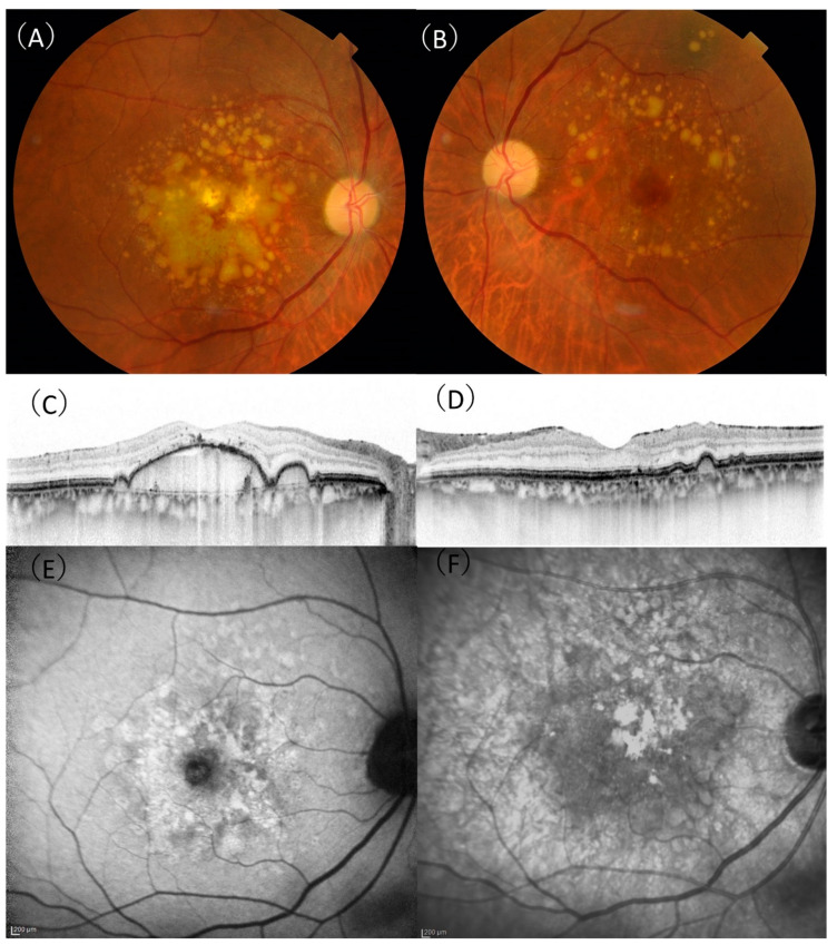
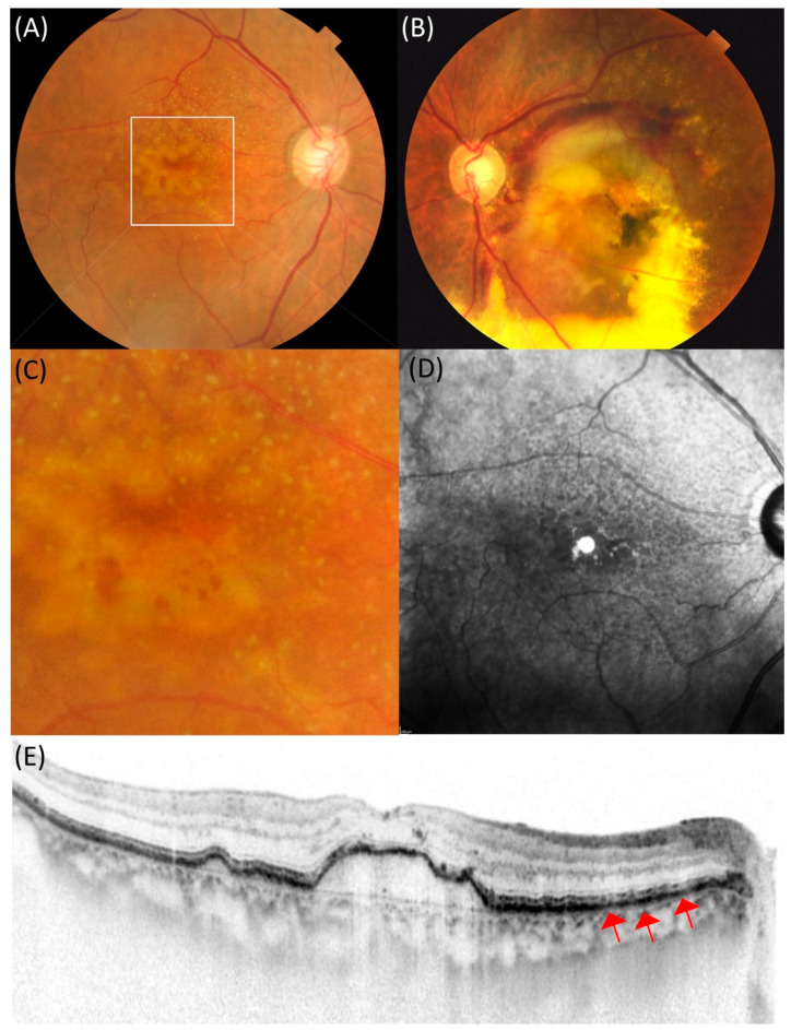
Few studies report drusenoid pigment epithelial detachment (DPED) in Asians. In this multicenter study, we report the clinical and genetic characteristics of 76 patients with DPED, and, for comparison, 861 patients with exudative age-related macular degeneration (AMD) were included. On the initial presentation, the mean best-corrected visual acuity was 0.087 ± 0.17 (logMAR unit), and mean DPED height and width were 210 ± 132 and 1633 ± 1114 µm, respectively. Fifty-one (67%) patients showed macular neovascularization in the contralateral eye. The risk allele frequency of both ARMS2 A69S and CFH I62V was significantly higher in DPED than in typical AMD and polypoidal choroidal vasculopathy (PCV) (ARMS2 A69S risk allele frequency: DPED 77% vs. typical AMD 66% vs. PCV 57%, CFH I62V risk allele frequency: DPED 87% vs. typical AMD 73% vs. PCV 73%), although the risk allele frequency of both genes was similar between the DPED group and retinal angiomatous proliferation (RAP) group (ARMS2 A69S: p = 0.32, CFH I62V, p = 0.11). The prevalence of reticular pseudodrusen (RPD) was highest in RAP (60%), followed by DPED (22%), typical AMD (20%), and PCV (2%). Although the prevalence of RPD differs between DPED and RAP, these entities share a similar genetic background in terms of ARMS2 and CFH genes.
Keywords: ARMS2; CFH; drusenoid pigment epithelial detachment; reticular pseudodrusen.
Conflict of interest statement
The authors declare no conflict of interest.
Figures
References
-
- Balaratnasingam C., Yannuzzi L.A., Curcio C.A., Morgan W.H., Querques G., Capuano V., Souied E., Jung J., Freund K.B. Associations Between Retinal Pigment Epithelium and Drusen Volume Changes During the Lifecycle of Large Drusenoid Pigment Epithelial Detachments. Investig. Opthalmol. Vis. Sci. 2016;57:5479–5489. doi: 10.1167/iovs.16-19816. - DOI - PMC - PubMed
-
- Cukras C., Agrón E., Klein M.L., Ferris F.L., 3rd, Chew E.Y., Gensler G., Wong W.T. Natural History of Drusenoid Pigment Epithelial Detachment in Age-Related Macular Degeneration: Age-Related Eye Disease Study Report No. 28. Ophthalmology. 2010;117:489–499. doi: 10.1016/j.ophtha.2009.12.002. - DOI - PMC - PubMed
-
- Yu J.J., Agrón E., Clemons T.E., Domalpally A., Van Asten F., Keenan T.D., Cukras C., Chew E.Y., Ferris F.L., SanGiovanni J.P., et al. Natural History of Drusenoid Pigment Epithelial Detachment Associated with Age-Related Macular Degeneration. Ophthalmology. 2019;126:261–273. doi: 10.1016/j.ophtha.2018.08.017. - DOI - PMC - PubMed
MeSH terms
LinkOut - more resources
Full Text Sources
Other Literature Sources
Medical
Miscellaneous
