. 2021 Apr 15;26(8):2282.
doi: 10.3390/molecules26082282.

Ursolic Acid Lactone Obtained from Eucalyptus tereticornis Increases Glucose Uptake and Reduces Inflammatory Activity and Intracellular Neutral Fat: An In Vitro Study

Affiliations

Ursolic Acid Lactone Obtained from Eucalyptus tereticornis Increases Glucose Uptake and Reduces Inflammatory Activity and Intracellular Neutral Fat: An In Vitro Study

Norman Balcazar et al. Molecules. .

Abstract

Obesity has a strong relationship to insulin resistance and diabetes mellitus, a chronic metabolic disease that alters many physiological functions. Naturally derived drugs have aroused great interest in treating obesity, and triterpenoids are natural compounds with multiple biological activities and antidiabetic mechanisms. Here, we evaluated the bioactivity of ursolic acid lactone (UAL), a lesser-known triterpenoid, obtained from Eucalyptus tereticornis. We used different cell lines to show for the first time that this molecule exhibits anti-inflammatory properties in a macrophage model, increases glucose uptake in insulin-resistant muscle cells, and reduces triglyceride content in hepatocytes and adipocytes. In 3T3-L1 adipocytes, UAL inhibited the expression of genes involved in adipogenesis and lipogenesis, enhanced the expression of genes involved in fat oxidation, and increased AMP-activated protein kinase phosphorylation. The range of biological activities demonstrated in vitro indicates that UAL is a promising molecule for fighting diabetes.

Keywords: adipocytes; diabetes; hepatocytes; macrophages; myocytes; ursolic acid lactone.


Conflict of interest statement

The authors declare no conflict of interest.

Figures
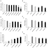
Figure 1
Figure 1
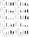
Effect of ursolic acid lactone (UAL) on cell viability, oxidative burst, and the expression of pro-inflammatory genes in activated J774A.1 macrophages. (A) J774A.1 cells were activated for 18 h and treated with various doses (0–200 μg/mL) of UAL. Cell viability was measured by the MTT assay after 6 h of treatment. The percentage of viable cells was calculated by defining the cell viability without treatment as 100%. Values are expressed as mean ± SEM of three independent experiments each one performed in triplicate. (B) J774A.1 cells were activated for 18 h and treated with various doses (0–100 μg/mL) of UAL. Cell viability was measured by 7-AAD fluorometric assay after 6 h of treatment. The percentage of viable cells was calculated by defining the cell viability without treatment as 100%. Values are expressed as mean ± SEM of three independent experiments each one performed in duplicate. (C) J774A.1 cells were activated for 18 h and treated with various doses (0–100 μg/mL) of UAL. Oxidative burst was quantified by flow cytometry using Dihydrorhodamine 123 (DHR) as qualitative marker of intracellular reactive oxygen species (ROS) after 6 h of treatment. PMA was used as positive control. Values are expressed as mean ± SEM of three independent experiments each one performed in duplicate. Relative expression of (D) Il1B, (E) Il6 and (F) Tnf mRNA transcripts is shown. J774A.1 cells were activated for 18 h and treated for 6 h with UAL. Metformin was used as positive control. Values are expressed as mean ± SEM of three independent experiments, normalized to the cyclophilin β gene expression. * p < 0.05, ** p < 0.01, *** p < 0.001, compared with LPS + INF-γ (activated cells) (ANOVA with Dunnett’s post hoc test).
Figure 2
Figure 2
Effect of UAL on cell viability and glucose uptake in C2C12 cells. (A) C2C12 cells were treated with various doses (0–50 μg/mL) of UAL. Cell viability was measured by the MTT assay after 4 h of treatment. The percentage of viable cells was calculated by defining the cell viability without treatment as 100%. Values are expressed as mean ± SEM of three independent experiments each one performed in triplicate. (B) Non-insulin-resistant C2C12 cells were treated with various doses (8–50 μg/mL) of UAL. Insulin (Ins) and metformin (Met) were used as positive controls. Values are expressed as mean ± SEM of three independent experiments each one performed in triplicate. (C) Insulin-resistant C2C12 cells were treated with various doses (8–50 μg/mL) of UAL. Insulin and metformin were used as controls. Values are expressed as mean ± SEM of three independent experiments each one performed in triplicate. * p < 0.05, *** p < 0.001 compared with CONTROL; # p < 0.05, ## p < 0.01 compared with CONTROL IR (ANOVA with Dunnett’s post hoc test).
Figure 3
Figure 3
Effect of UAL on cell viability and lipid accumulation in HepG2 hepatocytes. (A) HepG2 cells were treated with various doses (0–200 μg/mL) of UAL. Cell viability was measured by the MTT assay after 72 h of treatment. Values are expressed as mean ± SEM of three independent experiments each one performed in triplicate. (B) HepG2 cells were loaded with FFAs (oleic acid:palmitic acid/2:1) and treated with UAL for 72 h. Metformin was used as positive control. HepG2 cells were stained with oil red and lipid accumulation was quantified. Values are expressed as mean ± SEM of four independent experiments each one performed in triplicate. * p < 0.05, ** p < 0.05, *** p < 0.001 compared with FFA group (ANOVA with Dunnett´s post hoc test).
Figure 4
Figure 4
Effect of UAL on cell viability and oxidative burst in differentiated 3T3-L1 adipocytes. (A) 3T3-L1 cells were differentiated and treated with various doses (0–100 μg/mL) of UAL. Cell viability was measured by the MTT assay after 7 days of treatment. Values are expressed as mean ± SEM of three independent experiments each one performed in triplicate. (B) 3T3-L1 cells were differentiated and treated with various doses (0–50 μg/mL) of UAL. Cell viability was measured by 7-AAD fluorometric assay after 7 days of treatment. Values are expressed as mean ± SEM of three independent experiments each one performed in duplicate. (C) 3T3-L1 cells were differentiated and treated with various doses (0–50 μg/mL) of UAL. Oxidative burst was quantified by flow cytometry using Dihydrorhodamine 123 (DHR) as qualitative marker of intracellular reactive oxygen species (ROS) after 72 h of treatment. PMA was used as positive control. Values are expressed as mean ± SEM of three independent experiments each one performed in duplicate. ** p < 0.01, *** p < 0.001 compared with DIFFERENTIATED (ANOVA with Dunnett´s post hoc test).
Figure 5
Figure 5
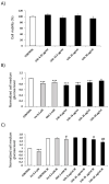
Effect of UAL on lipid accumulation in 3T3-L1 and protein expression in 3T3-L1 adipocytes. 3T3-L1 cells were differentiated and treated with UAL for 7 days. Metformin was used as positive control. (A) Photographs of 3T3-L1 adipocytes before and after Oil Red O staining. (B) 3T3L-1 cells were stained with oil red and lipid accumulation was quantified. Values are expressed as mean ± SEM of four independent experiments each one performed in triplicate. (C) Amount of adiponectin, and (D) amount of leptin present in the supernatant of the cells after 7 days of treatment. Values are expressed as mean ± SEM of three independent experiments each one performed in duplicate. * p < 0.05, ** p < 0.01, *** p < 0.001 vs. DIFFERENTIATED (ANOVA with Dunnett´s post hoc test).
Figure 6
Figure 6
Effect of UAL on the expression of functional genes in 3T3-L1 cells. 3T3-L1 cells were differentiated for 4 days and treated with UAL for 24 h. Metformin was used as positive control. Relative expression of Pparg (A), Cebpa (B), Adipoq (C), Ppara (D), Srebf1 (E), Acaca (F), Fasn (G) Lep (H), Ppargc1a (I), Ucp1 (J) mRNA transcripts is shown. Values are expressed as mean ± SEM of three independent experiments. * p < 0.05, ** p < 0.01, *** p < 0.001 compared with DIFFERENTIATED (ANOVA with Dunnett´s post hoc test).
Figure 7
Figure 7
Effect of UAL on differentiated 3T3-L1 mitochondria and cellular AMPK expression. 3T3-L1 cells were differentiated and treated with UAL for 72 h. (A) Mitochondrial membrane potential was determined by quantification of Mito tracker-loaded cells fluorescence. Values are expressed as mean ± SEM of three independent experiments each one performed in duplicate. (B) ADP/ATP ratio in 3T3-L1 adipocytes treated with UAL. Values are expressed as mean ± SEM of three independent experiments each one performed in triplicate (C) Representative western blots of phosphorylated AMPK in differentiated 3T3-L1 cultures treated with UAL. (D) Quantitative analysis of the immunoblots of p-AMPK normalized with total AMPK. Values are expressed as mean ± SEM of three independent experiments. * p < 0.05, ** p < 0.01 compared with DIFFERENTIATED (ANOVA with Dunnett´s post hoc test).
Figure 8
Figure 8
Chromatogram of Eu extract. Chromatographic profile of E. tereticornis leaves extract. UAL: Ursolic acid lactone.

References

    1. de Freitas Junior L.M., de Almeida E.B. Medicinal plants for the treatment of obesity: Ethnopharmacological approach and chemical and biological studies. Am. J. Transl. Res. 2017;9:2050. - PMC - PubMed
    1. International Diabetes Federation . IDF Diabetes Atla. 9th ed. Volume 2019 International Diabetes Federation; Brussels, Belgium: 2019.
    1. Schnurr T.M., Jakupović H., Carrasquilla G.D., Ängquist L., Grarup N., Sørensen T.I.A., Tjønneland A., Overvad K., Pedersen O., Hansen T., et al. Obesity, unfavourable lifestyle and genetic risk of type 2 diabetes: A case-cohort study. Diabetologia. 2020 doi: 10.1007/s00125-020-05140-5. - DOI - PubMed
    1. Smith U., Kahn B.B. Adipose tissue regulates insulin sensitivity: Role of adipogenesis, de novo lipogenesis and novel lipids. J. Intern. Med. 2016 doi: 10.1111/joim.12540. - DOI - PMC - PubMed
    1. Ríos J.L., Francini F., Schinella G.R. Natural products for the treatment of type 2 diabetes mellitus. Planta Med. 2015;81:975–994. doi: 10.1055/s-0035-1546131. - DOI - PubMed

LinkOut - more resources