. 2021 Apr 15;9(4):389.
doi: 10.3390/vaccines9040389.

A Simplified SARS-CoV-2 Pseudovirus Neutralization Assay

Affiliations

A Simplified SARS-CoV-2 Pseudovirus Neutralization Assay

Gaetano Donofrio et al. Vaccines (Basel). .

Abstract

COVID-19 is an ongoing pandemic caused by the highly infectious coronavirus SARS-CoV-2 that is engaging worldwide scientific research to find a timely and effective eradication strategy. Great efforts have been put into anti-COVID-19 vaccine generation in an effort to protect the world population and block SARS-CoV-2 spread. To validate the protective efficacy of the vaccination campaign and effectively control the pandemic, it is necessary to quantify the induction of neutralizing antibodies by vaccination, as they have been established to be a correlate of protection. In this work, a SARS-CoV-2 pseudovirus neutralization assay, based on a replication-incompetent lentivirus expressing an adapted form of CoV-2 S protein and an ACE2/TMPRSS2 stably expressing cell line, has been minimized in terms of protocol steps without loss of accuracy. The goal of the present simplified neutralization system is to improve SARS-CoV-2 vaccination campaign by means of an easy and accessible approach to be performed in any medical laboratory, maintaining the sensitivity and quantitative reliability of classical serum neutralization assays. Further, this assay can be easily adapted to different coronavirus variants by simply modifying the pseudotyping vector.

Keywords: COVID-19; SARS-CoV-2; neutralization assay; neutralizing antibody; pseudovirus.


Conflict of interest statement

No potential conflict of interest was reported by the authors.

Figures

Figure 1
Figure 1
CoV-2 S expression in HEK293T cells. (A) Diagram (not to scale) of pLV-CMV-(S-ΔRS-HA)-IRES-Puro-WPRE transfer vector, where functional elements of the construct are indicated and highlighted with different colors. RSVp (Rous sarcoma virus enhancer/promoter), 5′LTRΔU3 (truncated HIV-1 5′ long terminal repeat), Ψ (HIV-1 packaging signal), RRE (HIV-1 REV response element), cPPT (central polypurine tract), CMV (human cytomegalovirus immediate early enhancer/promoter), Kozak (Kozak’s translation initiation sequence), S-ΔRS-HA (human codon usage adapted, HA-tagged and retention-signal-deleted CoV-2 S ORF), IRES (encephalomyocarditis virus internal ribosomal entry site), Puro (puromycin-resistant gene ORF), WPRE (woodchuck hepatitis virus posttranslational regulatory element), 3′LTRΔU3 (truncated HIV-1 3′ long terminal repeat; self-inactivation), SV40 early pA (simian virus 40 early polyadenylation signal), Amp (ampicillin resistance gene, comprising the promoter and ORF) and pUC ori (high-copy-number pUC origin of replication). (B) Western immunoblotting of pLV-CMV-(S-ΔRS-HA)-IRES-Puro-WPRE transfected HEK293T cell protein extracts (S-ΔRS-HA; 40 and 20 µg) and pEGFP-C1 transfected HEK293T cell protein extract (GFP; 40 µg) employed as a negative control (-). Cross-reactive bands in both lanes indicate the loading. (C) Representative microscopic image (phase contrast, fluorescence, and merged fields; 10X) of syncytia, generated by cotransfection of HEK/ACE2/TMPRRS2/Puro cells with pLV-CMV-(S-ΔRS-HA)-IRES-Puro-WPRE construct and PEGFP-C1. (D) Representative microscopic image (phase contrast, fluorescence, and merged fields; 10X) of syncytia, generated by cocultivation of HEK/ACE2/TMPRRS2/Puro cells with pLV-CMV-(S-ΔRS-HA)-IRES-Puro-WPRE and pEGFP-C1 cotransiently transfected HEK293T cells.
Figure 2
Figure 2
Pseudovirus assembly and transduction. (A) Representative images (phase contrast and fluorescence; 10X) of HEK/ACE2/TMPRRS2/Puro cells, at different numbers (1000 and 4000), transduced with different amounts (50, 20, and 10 µL) of supernatant containing pseudovirus. (B) ChemiDoc and (C) IVIS luminometric detection of HEK/ACE2/TMPRRS2/Puro cells, at different numbers (1000, 2000, 3000, 4000, 5000, and 6000), transduced with different amounts (50, 20, and 10 µL) of supernatant containing pseudovirus (S pseudovirus). Negative control was established with HEK293T cells, at different numbers (1000, 2000, 3000, 4000, 5000, and 6000) and transduced with 50 µL of the same supernatant containing pseudovirus (S pseudovirus). (D) Representative images of syncytia (phase contrast and fluorescence of the same field; 10X), generated by pseudovirus-transduced HEK/ACE2/TMPRRS2/Puro cells. (E) Representative images of a syncytium (phase contrast; 10X) generated by pseudovirus-transduced HEK/ACE2/TMPRRS2/Puro cells and stained with hematoxylin and eosin (H&E staining).
Figure 3
Figure 3
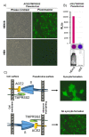
Pseudovirus generation with ACE2 and TMPRSS2. (A) Representative image (phase contrast and fluorescence of the same field; 10X) of HEK/S-ΔRS-HA/Puro cells (HEK/S) and HEK293T cells (HEK) transduced with ACE2/TMPRSS2 pseudovirus. (B) Representative 25 cm2 flasks of HEK/S-ΔRS-HA/Puro cells (HEK/S) and HEK293T cells (HEK) transduced with ACE2/TMPRSS2 pseudovirus and exposed to the ChemiDoc apparatus. Luminometric quantification of the lysate of the same cells, expressed as relative luciferase units (RLUs) and colorimetrically visualized (violet and blue spots) by the luminometer software, during and after reading. (C) Drawing explaining the lack of syncytia generation by ACE2/TMPRSS2 pseudovirus.
Figure 4
Figure 4
Representative pseudovirus neutralization assay. Six PCR and ELISA CoV-2 negative patients’ representative sera (1 to 6), three PCR and ELISA CoV-2 positive patients’ sera (7 to 9), and two pre-pandemic sera (collected in 1998; 10 and 11) were tested. A further negative control was established without serum (12), which was substituted with complete medium. (A) Histogram obtained from luminometric detection of transduced cells with pseudovirus. Relative luciferase units (RLUs) were compared and normalized to those derived from wells where pseudovirus was added in the absence of sera (100%). Neutralization titer 50 (NT50) was expressed as the maximal dilution of the sera where the reduction of the signal was ≥50%. In this specific example, the NT50/25 µL of serum 7 is 1:256, that of serum 8 is 1:128, and that of serum 9 is 1:64. However, because the titer was measured at 25 µL, the apparent NT50 has to be multiplied by 40 to obtain the normalized NT50/mL. Therefore, the NT50/mL of the sera 7 is 1:10,256, that of the serum 8 is 1:5120, and that of the serum 9 is 1:2560. Bar colors indicate sera dilution. (B) Colorimetric visualization of the RLUs displayed by the luminometer software, during and after reading.

References

    1. Han Q., Lin Q., Jin S., You L. Coronavirus 2019-nCoV: A brief perspective from the front line. J. Infect. 2020;80:373–377. doi: 10.1016/j.jinf.2020.02.010. - DOI - PMC - PubMed
    1. Callaway E. The race for coronavirus vaccines: A graphical guide. Nature. 2020;580:576–577. doi: 10.1038/d41586-020-01221-y. - DOI - PubMed
    1. Poland G.A., Ovsyannikova I.G., Kennedy R.B. SARS-CoV-2 immunity: Review and applications to phase 3 vaccine candidates. Lancet. 2020;396:1595–1606. doi: 10.1016/S0140-6736(20)32137-1. - DOI - PMC - PubMed
    1. Iacob S., Iacob D.G. SARS-CoV-2 Treatment Approaches: Numerous Options, No Certainty for a Versatile Virus. Front. Pharmacol. 2020;11:1224. doi: 10.3389/fphar.2020.01224. - DOI - PMC - PubMed
    1. Li G., De Clercq E. Therapeutic options for the 2019 novel coronavirus (2019-nCoV) Nat. Rev. Drug Discov. 2020;19:149–150. doi: 10.1038/d41573-020-00016-0. - DOI - PubMed