. 2021 Apr 12;11(4):985.
doi: 10.3390/nano11040985.

A Label-Free DNA-Immunosensor Based on Aminated rGO Electrode for the Quantification of DNA Methylation

Affiliations

A Label-Free DNA-Immunosensor Based on Aminated rGO Electrode for the Quantification of DNA Methylation

Mina Safarzadeh et al. Nanomaterials (Basel). .

Abstract

In this work, we developed a sandwich DNA-immunosensor for quantification of the methylated tumour suppressor gene O-6-methylguanine-DNA methyltransferase (MGMT), which is a potential biomarker for brain tumours and breast cancer. The biosensor is based on aminated reduced graphene oxide electrode, which is achieved by ammonium hydroxide chemisorption and anti-5-methylcytosine (anti-5mC) as a methylation bioreceptor. The target single-strand (ss) MGMT oligonucleotide is first recognised by its hybridisation with complementary DNA to form double-stranded (ds) MGMT, which is then captured by anti-5mC on the electrode surface due to the presence of methylation. Raman spectroscopy, X-ray photoelectron spectroscopy (XPS) and Scanning electron microscopy (SEM) techniques were used to characterise the electrode surface. Cyclic voltammetry (CV) and differential pulse voltammetry (DPV) techniques were used for electrochemical measurements. Under optimised conditions, the proposed biosensor is able to quantify a linear range of concentrations of the MGMT gene from 50 fM to 100 pM with a limit of detection (LOD) of 12 fM. The sandwich design facilitates the simultaneous recognition and quantification of DNA methylation, and the amination significantly improves the sensitivity of the biosensor. This biosensor is label-, bisulfite- and PCR-free and has a simple design for cost-efficient production. It can also be tailor-made to detect other methylated genes, which makes it a promising detection platform for DNA methylation-related disease diagnosis and prognosis.

Keywords: MGMT gene; NH2 chemisorption; amination; quantification of DNA methylation; reduced graphene oxide (rGO).


Conflict of interest statement

The authors declare no conflict of interest.

Figures

Figure A1
Figure A1
SEM images of a bare rGO electrode (A), an aminated rGO electrode (B) and aminated electrodes after incubation in the antibody (C).
Figure A2
Figure A2
Detection of 100 (purple) and 1000 (blue) pM of the target gene using either the antibody or antibody conjugated to protein G, using both CV (A) and DPV (B) techniques.
Figure A3
Figure A3
Detection of 100 (purple) and 1000 (blue) pM of the target gene with various bovine serum albumin (BSA) optimisation times (5, 15 and 30 min), using both CV (A) and DPV (B) techniques.
Figure A4
Figure A4
Detection of 100 (purple) and 1000 (blue) pM of the target gene of the target gene with various antibody incubation times (1, 2, 3, 4, 5, 6 and 8 h) using both CV (A) and DPV (B) techniques.
Figure A5
Figure A5
Detection of 100 (purple) and 1000 (blue) pM of the target gene of the target gene with various antigen incubation times (30, 60, 90 and 120 min) using both CV (A) and DPV (B) techniques.
Figure A6
Figure A6
Comparison of the responses obtained from various targets (methylated, non-methylated and blank) using the proposed biosensor. Normalized CV peak current for different samples measured on three different replicas (A). CV (B) and DPV (C) voltammograms of one replica for each sample.
Figure A7
Figure A7
CV voltammograms of the aminated electrode under various scan rates from 0.025 V/s to 0.3 V/s (0.025, 0.05, 0.075, 0.1, 0.125, 0.15, 0.175, 0.2, 0.225, 0.275 and 0.3 V/s) (A). Anodic and Cathodic peaks as a function of the square root of the scan rate (B) and the scan rate (C).
Figure 1
Figure 1
Raman spectra obtained from a bare reduced graphene oxide (rGO) electrode and rGO electrode incubated in ammonium hydroxide. Both the G (1578 cm1) and D (1340 cm1) bands decreased and broadened after rGO amination while the ID/IG ratio increased from 0.6 to 0.7.
Figure 2
Figure 2
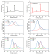
XPS spectra of rGO and aminated rGO electrodes. Survey scan of a bare rGO electrode (a) and an aminated electrode (b). N1s high resolution spectra of bare rGO (c) and aminated rGO (d) electrodes and C1s high resolution spectra of bare rGO (e) and aminated rGO (f) electrodes.
Figure 3
Figure 3
Schematic of the possible surface reactions that may occur on the rGO electrode after incubation in ammonium hydroxide. These reactions would lead to the presence of amine functional groups on the surface.
Figure 4
Figure 4
Schematic display of the developed method for the quantification of O-6-methylguanine-DNA methyltransferase (MGMT) oligonucleotide (a). Electrodes were incubated in ammonium hydroxide and were kept in a vacuum for further use. Cyclic voltammetry (CV) (b) and differential pulse voltammetry (DPV) (c) characteristics of the sensor after each assembly steps in 10 mM K3[Fe(CN)6] containing 1 M KCl.
Figure 5
Figure 5
Calibration curves constructed with normalised peak currents of DPV responses as a function of the logarithm of the concentration of target ssDNA (red) and dsDNA (black). For both targets, the current increases with increases in concentration. Error bars are the standard deviation of three replicates.

References

    1. Davis C.D., Uthus E.O. DNA methylation, cancer susceptibility, and nutrient interactions. Exp. Biol. Med. 2004;229:988–995. doi: 10.1177/153537020422901002. - DOI - PubMed
    1. Syedmoradi L., Esmaeili F., Norton M.L. Towards DNA methylation detection using biosensors. Analyst. 2016;141:5922–5943. doi: 10.1039/C6AN01649A. - DOI - PubMed
    1. Bhattacharjee R., Moriam S., Umer M., Nguyen N.T., Shiddiky M.J. DNA methylation detection: Recent developments in bisulfite free electrochemical and optical approaches. Analyst. 2018;143:4802–4818. doi: 10.1039/C8AN01348A. - DOI - PubMed
    1. Kurdyukov S., Bullock M. DNA methylation analysis: Choosing the right method. Biology. 2016;5:3. doi: 10.3390/biology5010003. - DOI - PMC - PubMed
    1. Shanmuganathan R., Basheer N.B., Amirthalingam L., Muthukumar H., Kaliaperumal R., Shanmugam K. Conventional and nanotechniques for DNA methylation profiling. J. Mol. Diagn. 2013;15:17–26. doi: 10.1016/j.jmoldx.2012.06.007. - DOI - PubMed

LinkOut - more resources