. 2021 Apr 12;22(8):3968.
doi: 10.3390/ijms22083968.

Rapamycin Alleviates 2,3,7,8-Tetrachlorodibenzo-p-dioxin-Induced Aggravated Dermatitis in Mice with Imiquimod-Induced Psoriasis-Like Dermatitis by Inducing Autophagy

Affiliations

Rapamycin Alleviates 2,3,7,8-Tetrachlorodibenzo-p-dioxin-Induced Aggravated Dermatitis in Mice with Imiquimod-Induced Psoriasis-Like Dermatitis by Inducing Autophagy

Hye Ran Kim et al. Int J Mol Sci. .

Abstract

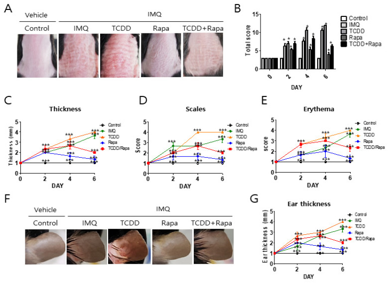
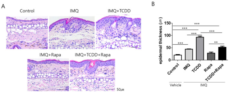
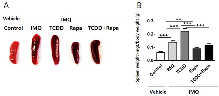
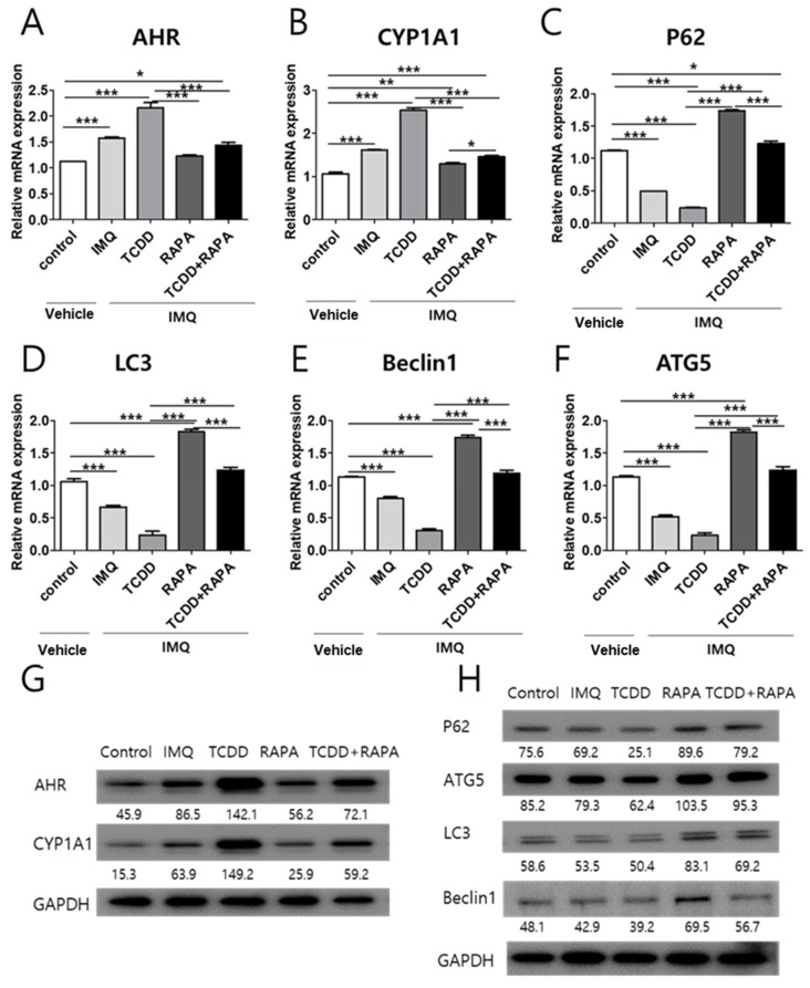
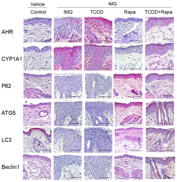
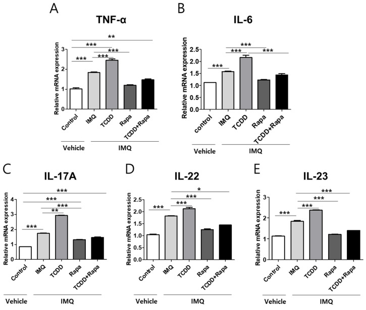
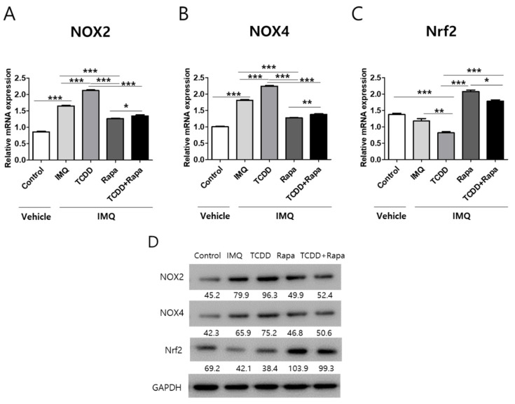
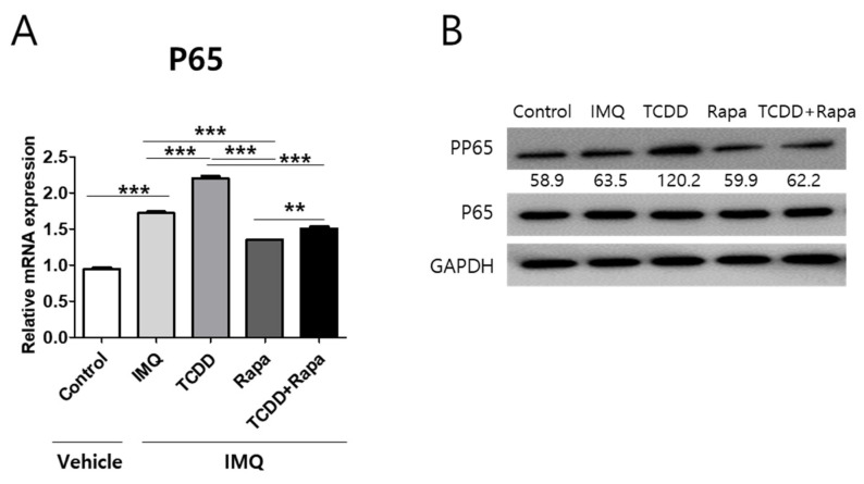
Recently, the mTOR signaling has emerged as an important player in the pathogenesis of psoriasis. We previously found that 2,3,7,8-tetrachlorodibenzo-p-dioxin (TCDD)-induced psoriatic skin inflammation was related to the inhibition of autophagy in keratinocytes. However, the effects and detailed molecular mechanisms of the mTOR inhibitor rapamycin and TCDD on psoriasis in vivo remain to be elucidated. In this study, we aimed to evaluate the effects of rapamycin and TCDD on skin lesions in imiquimod (IMQ)-induced psoriasis using a mouse model. TCDD aggravated skin inflammation in an IMQ-induced psoriatic mouse model. Furthermore, TCDD increased the expression of aryl hydrocarbon receptor (AHR), CYP1A1, proinflammatory cytokines, oxidative stress markers (NADPH oxidase (Nox) 2, Nox4), and phosphorylated P65NF-ĸB, whereas the expression of autophagy-related factors and the antioxidant marker nuclear factor-erythroid 2-related factor 2 (NRF2) decreased. Rapamycin reduced the aggravated skin inflammation induced by TCDD and restored TCDD-induced autophagy suppression and the increase of AHR expression, oxidative stress, and inflammatory response in the skin lesions of a psoriatic mouse model. In conclusion, we demonstrated that rapamycin alleviates TCDD-induced aggravated dermatitis in mice with imiquimod-induced psoriasis-like dermatitis through AHR and autophagy modulation.

Keywords: 2,3,7,8-tetrachlorodibenzo-p-dioxin; aryl hydrocarbon receptor; autophagy; psoriasis; rapamycin.


Conflict of interest statement

The authors declare no conflict of interest.

Figures

Figure 1
Figure 1
Study procedures. C57BL/6 mice were treated on the dorsal skin and right ear pinna for 4 days with five different treatment regimens as described in the Section 4. C57BL/6 mice were treated daily with vehicle control, IMQ, IMQ + TCDD, IMQ + Rapa, or IMQ + TCDD + Rapa applied on their shaved back skin and right ears. IMQ: imiquimod, Rapa: rapamycin.
Figure 2
Figure 2
The effects of TCDD, rapamycin, and cotreatment with both on the clinical severity of imiquimod-induced psoriatic mice. C57BL/6 mice were treated daily with vehicle control, IMQ, IMQ + TCDD, IMQ + Rapa, or IMQ + TCDD + Rapa applied on their shaved back skin and right ears (n = 5 per treatment group). (A) Macroscopic presentation of dorsal skin of mice from the 5 treatment regimens. (B) The severity of inflammation on the back was assessed using a scoring system similar to the human Psoriasis Area and Severity Index (PASI) score. Thickness, scaling, and erythema of the back skin were scored “blindly” on a scale from 0 to 3 as follows: 0: none; 1: slight; 2: moderate; and 3: severe. (CE) Detailed clinical disease score of thickness, scales, and erythema. Data represent mean ± standard deviation (SD). Statistical significance was determined by two-way repeated measures analysis of variance (ANOVA) with Tukey’s test. * p < 0.05; *** p < 0.001 compared with controls. (F) Macroscopic presentation of the right ear of mice from the 5 treatment regimens. (G) Assessment of ear skin thickness of the mice measured throughout the experiment. Statistical significance was determined by two-way repeated measures analysis of variance (ANOVA) with Tukey’s test. * p < 0.05; *** p < 0.001 compared with controls. IMQ: imiquimod, Rapa: rapamycin.
Figure 3
Figure 3
Histological changes after treatment with TCDD, rapamycin, or both in an IMQ-induced psoriasis mouse model. (A) Hematoxylin and eosin (H&E) staining of representative images from each group (dorsal skin). Bars represent 50 μm. (B) Evaluation of epidermal thickness in dorsal skin. The epidermal thickness was measured from the bottom of the rete ridge to the bottom of the stratum corneum; the mean was calculated from the measurements in four fields at ×100 magnification. Data represent mean ± standard deviation (SD). Statistical significance was determined by one-way repeated measures analysis of variance (ANOVA) with Dunnett’s test. ** p < 0.01; *** p < 0.001. IMQ: imiquimod, Rapa: rapamycin.
Figure 4
Figure 4
The spleen size and weight of mice from 5 study groups. (A) Spleens were prepared from each mouse and weighed. The spleen shown is from 1 representative experiment with n = 5 per treatment group. (B) The spleens from mice were isolated and weighed, and the spleen weight index was calculated as organ weight (milligram, mg) per gram (g) of mouse body weight. All data are presented as mean ± standard deviation (SD). Statistical differences were determined by one-way repeated measures analysis of variance (ANOVA) with Dunnett’s test. ** p < 0.01; *** p < 0.001. IMQ: imiquimod, Rapa: rapamycin.
Figure 5
Figure 5
mRNA and Western blot analysis of AHR, CYP1A1, and autophagy-related factor expression changes in skin tissue with treatment with vehicle control, IMQ, IMQ + TCDD, and IMQ + Rapa, or IMQ + TCDD + Rapa. RNA and protein were extracted from the back skin. (AF) qPCR and (G,H) Western blot analysis of AHR, CYP1A1, P62, ATG5, LC3, and Beclin1 expressions in mice treated with 5 different regimens (n = 5 per treatment group). qPCR data represent the mean ± standard deviation (SD) of three independent experiments (each performed in duplicate). Statistical differences were determined by one-way analysis of variance (ANOVA) followed by post hoc Dunnett’s test. * p < 0.05; ** p < 0.01; *** p < 0.001. Western blot normalization was based on GAPDH. Western blot data are representative of three independent experiments. IMQ: imiquimod, Rapa: rapamycin.
Figure 6
Figure 6
Immunohistochemical analysis of AHR, CYP1A1, and autophagy-related factors in skin tissue of mice from 5 different treatment regimens with vehicle control, IMQ, IMQ + TCDD, and IMQ + Rapa, or IMQ + TCDD + Rapa. Immunohistochemical staining with the indicated antibodies is shown from 1 representative mouse from each group (n = 5). Scale bars represent 50 μm. IMQ: imiquimod, Rapa: rapamycin.
Figure 7
Figure 7
The effects of TCDD, rapamycin, or cotreatment of both on proinflammatory cytokine mRNA expression changes in the skin tissues of a psoriasis mouse model. (AE) qPCR analysis of TNF-α, IL-6, IL-17A, IL-22, and IL-23 was performed in skin tissue from 5 different study groups (n = 5 per group). Data represent mean ± SD of three independent experiments (each performed in duplicate). Statistical differences were determined by one-way analysis of variance (ANOVA) with Dunnett’s test. * p < 0.05; ** p < 0.01; *** p < 0.001. Rapa: rapamycin; IMQ: imiquimod.
Figure 8
Figure 8
The effects of TCDD and rapamycin on the expression of oxidative stress-related factors. mRNA and protein expressions change in mouse skin tissue after treatment with vehicle control, IMQ, IMQ + TCDD, and IMQ + Rapa, or IMQ + TCDD + Rapa. (AC) qPCR and (D) Western blot analyses of NOX2, NOX4, and Nrf2 expressions in mice treated with 5 different regimens (n = 5 per group). qPCR data represent the mean ± SD of three independent experiments (each performed in duplicate). Statistical differences were determined by one-way analysis of variance (ANOVA) with Dunnett’s post hoc test. * p < 0.05; ** p < 0.01; *** p < 0.001. Western blot normalization was based on GAPDH. Western blot data are representative of three independent experiments. Rapa: rapamycin; IMQ: imiquimod.
Figure 9
Figure 9
The effects of TCDD, rapamycin, or a combination of both on P65 mRNA and phosphorylation of P65 expression changes in mouse skin tissue. (A) qPCR of P65 and (B) Western blot analysis of phosphorylation of P65 expression in mouse skin from 5 different treatment regimens (n = 5 per group). Data represent the mean ± SD of three independent experiments (each performed in duplicate). Statistical differences were determined by one-way analysis of variance (ANOVA) followed by post hoc Dunnett’s test. ** p < 0.01; *** p < 0.001. Western blot normalization was based on GAPDH. Western blot data are representative of three independent experiments. IMQ: imiquimod, Rapa: rapamycin.

Similar articles

Cited by

References

    1. Parisi R., Symmons D.P., Griffiths C.E., Ashcroft D.M. Global epidemiology of psoriasis: A systematic review of incidence and prevalence. J. Investig. Dermatol. 2013;133:377–385. doi: 10.1038/jid.2012.339. - DOI - PubMed
    1. Boehncke W.H., Schon M.P. Psoriasis. Lancet. 2015;386:983–994. doi: 10.1016/S0140-6736(14)61909-7. - DOI - PubMed
    1. Liu Y., Krueger J.G., Bowcock A.M. Psoriasis: Genetic associations and immune system changes. Genes Immun. 2007;8:1–12. doi: 10.1038/sj.gene.6364351. - DOI - PubMed
    1. Santus P., Rizzi M., Radovanovic D., Airoldi A., Cristiano A., Conic R., Petrou S., Pigatto P.D.M., Bragazzi N., Colombo D., et al. Psoriasis and Respiratory Comorbidities: The Added Value of Fraction of Exhaled Nitric Oxide as a New Method to Detect, Evaluate, and Monitor Psoriatic Systemic Involvement and Therapeutic Efficacy. BioMed Res. Int. 2018;2018:3140682. doi: 10.1155/2018/3140682. - DOI - PMC - PubMed
    1. Conic R.R., Damiani G., Schrom K.P., Ramser A.E., Zheng C., Xu R., McCormick T.S., Cooper K.D. Psoriasis and Psoriatic Arthritis Cardiovascular Disease Endotypes Identified by Red Blood Cell Distribution Width and Mean Platelet Volume. J. Clin. Med. 2020;9:186. doi: 10.3390/jcm9010186. - DOI - PMC - PubMed

MeSH terms

LinkOut - more resources