. 2021 Apr 12;22(8):3979.
doi: 10.3390/ijms22083979.

LeishIF4E-5 Is a Promastigote-Specific Cap-Binding Protein in Leishmania

Affiliations

LeishIF4E-5 Is a Promastigote-Specific Cap-Binding Protein in Leishmania

Rohit Shrivastava et al. Int J Mol Sci. .

Abstract

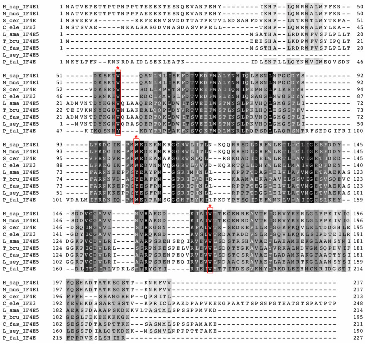
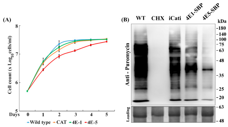
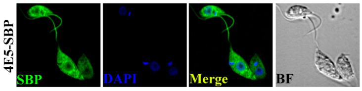
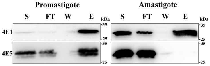
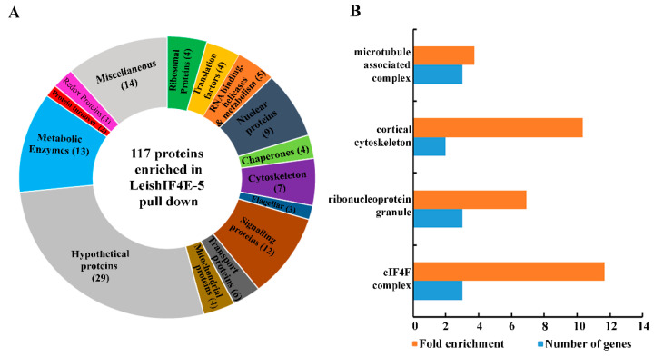
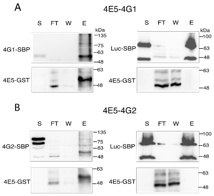
Leishmania parasites cycle between sand fly vectors and mammalian hosts, transforming from extracellular promastigotes that reside in the vectors' alimentary canal to obligatory intracellular non-motile amastigotes that are harbored by macrophages of the mammalian hosts. The transition between vector and host exposes them to a broad range of environmental conditions that induces a developmental program of gene expression, with translation regulation playing a key role. The Leishmania genome encodes six paralogs of the cap-binding protein eIF4E. All six isoforms show a relatively low degree of conservation with eIF4Es of other eukaryotes, as well as among themselves. This variability could suggest that they have been assigned discrete roles that could contribute to their survival under the changing environmental conditions. Here, we describe LeishIF4E-5, a LeishIF4E paralog. Despite the low sequence conservation observed between LeishIF4E-5 and other LeishIF4Es, the three aromatic residues in its cap-binding pocket are conserved, in accordance with its cap-binding activity. However, the cap-binding activity of LeishIF4E-5 is restricted to the promastigote life form and not observed in amastigotes. The overexpression of LeishIF4E-5 shows a decline in cell proliferation and an overall reduction in global translation. Immuno-cytochemical analysis shows that LeishIF4E-5 is localized in the cytoplasm, with a non-uniform distribution. Mass spectrometry analysis of proteins that co-purify with LeishIF4E-5 highlighted proteins involved in RNA metabolism, along with two LeishIF4G paralogs, LeishIF4G-1 and LeishIF4G-2. These vary in their conserved eIF4E binding motif, possibly suggesting that they can form different complexes.

Keywords: LeishIF4E-5; LeishIF4G; Leishmania; protein synthesis; translation regulation.


Conflict of interest statement

No conflict of interest.

Figures

Figure 1
Figure 1
Alignment of LeishIF4E-5 from L. amazonensis with its protozoan orthologs, with eIF4E-1 from mammals, with yeast eIF4E and C. elegans IFE-3. The alignment includes sequences of LeishIF4E-5 from Leishmania amazonensis (L_ama_IF4E5), Trypanosoma brucei (T_bru_IF4E5), Crithidia fasciculata (C_fas_IF4E5) and Leptomonas seymouri (L_sey_IF4E5). It also includes the sequences of the canonical eIF4E from Homo sapiens (H_sap_IF4E-1), Mus musculus (M_mus_IF4E-1) and Saccharomyces cerevisiae (S_cer_IF4E5) along with IFE-3 from C. elegans (C_ele_IFE3) and P. falciparum (P_fal_IF4E). All sequences were subjected to multiple sequence alignment to highlight their homologies. Conserved tryptophan residues involved in cap-binding are marked with asterisks. The multiple sequence alignment was performed using MAFFT, version 7. Sequence conservations were generated by Jalview and are highlighted in greyscale.
Figure 2
Figure 2
Episomal expression of LeishIF4E-5 affects global translation and growth. Wild type L. amazonensis, transgenic L. amazonensis promastigotes expressing chloramphenicol acetyltransferase (CAT), SBP-tagged LeishIF4E-5 and SBP-tagged LeishIF4E-1 were grown under normal conditions. (A) Cell counts were taken during 5 days. Cells expressing LeishIF4E-5 tagged with the streptavidin binding peptide, SBP (4E5-SBP), are shown in red, LeishIF4E-1-SBP cells are shown in green, cells expressing the CAT reporter are shown in orange and wild type cells are shown in blue. Cell growth is represented by log10 cells/mL against the number of days, taken in triplicates. Lines were drawn based on the mean values of the different experiments with standard errors. (B) Cells were incubated with 1 µg/mL puromycin for 20 min, extracted proteins were separated over 12% SDS-PAGE and subjected to Western analysis using specific antibodies against puromycin. A cycloheximide (CHX) control for complete translation inhibition is also shown. The different lanes contain equal protein loads, as shown by the Ponceau staining (bottom panel). The experiment was repeated three times and the densitometric analysis is given in Figure S3.
Figure 3
Figure 3
Non-uniform distribution of SBP-tagged LeishIF4E-5 in the cytoplasm and in foci surrounding the nucleus. Transgenic L. amazonensis promastigotes expressing SBP-tagged LeishIF4E-5 were fixed, permeabilized and processed for confocal microscopy. LeishIF4E-5-SBP was detected using a monoclonal antibody against the SBP tag and a secondary DyLight-labeled antibody (488 nm; green). Nuclear and kinetoplast DNA was stained using DAPI (blue). A bright field (BF) picture of the cells is on the right. The confocal analysis was repeated three times. A broad view of the field is given in Figure S4.
Figure 4
Figure 4
LeishIF4E-5 binds to the cap analog m7GTP only during the promastigote stage. Transgenic L. amazonensis promastigotes expressing SBP-tagged LeishIF4E-5 were lysed and incubated with m7GTP-agarose beads. The beads were washed and eluted using 200 μM free m7GTP. Samples from the eluted protein fractions and washes were precipitated by 10% trichloro-acetic acid (TCA). The gels were loaded with samples of the total supernatant (S, 2%), flow-through (FT, 2%), the wash (W, 50%) and the eluted fractions (E, 50%). The proteins were resolved over 12% SDS-PAGE and analyzed using specific antibodies against the SBP-tag to identify LeishIF4E-5 and antibodies raised against LeishIF4E-1. Similar results were obtained from three independent experiments. Densitometric analysis of the cap-binding activity in promastigotes is given in Figure S5.
Figure 5
Figure 5
Categorized proteins found enriched with LeishIF4E-5. SBP-LeishIF4E-5 and its associated proteins were pulled-down over streptavidin-Sepharose beads. Control pull downs were performed with cells expressing SBP-luciferase. The proteomic content of the pulled-down extracts was assessed by LC-MS/MS. Proteins were identified using the MaxQuant software and their enrichment as compared to the luciferase control was determined using the Perseus statistical tool, highlighting a log2 fold change of ≥1.6, with an adjusted p value (Padj) < 0.05. A total of 117 proteins were found to be significantly enriched in the interactome of SBP-LeishIF4E-5 that served as the bait protein. The proteins were categorized into groups according to their annotated functions. (A) The pie chart shows the relative distribution of proteins that were manually categorized into functional groups. The pie chart shows the relative abundance of each category, based on the summed intensities of the peptides that were used to identify the individual proteins. The numbers in brackets in the pie chart represent the number of proteins in each category. (B) Gene Ontology (GO) term enrichment by cellular components. The resulting GO terms were enriched by at least three-fold as compared to the gene sets encoded in the genome, with a p value ≤ 0.05.
Figure 6
Figure 6
Recombinant LeishIF4E-5-GST interacts directly with LeishIF4G-1 and LeishIF4G-2. Lysates of Leishmania cell lines expressing SBP-tagged LeishIF4G-1 or LeishIF4G-2 were bound to Streptavidin beads, followed by incubation with E. coli cell lysates expressing recombinant GST-tagged LeishIF4E-5. After several washes, the final elution was carried out with 5 mM Biotin. Leishmania cell line expressing SBP-tagged luciferase was used as a negative control. Aliquots obtained from the supernatant (S, 2%), the flow-through (FT, 2%), the wash (W, 25%) and the eluted fractions (E, 25%) were resolved over 12.5% SDS-PAGE, and subjected to Western blot analysis with specific monoclonal antibodies against the SBP and GST tags. (A) LeishIF4E-5-GST interacts directly with LeishIF4G-1. The top panels represent the blot developed with anti-SBP antibodies while the bottom panels represent the blot developed with anti-GST antibodies. (B) LeishIF4E-5-GST interacts directly with LeishIF4G-2-SBP. The top panels represent the blot developed with anti-SBP antibodies while the bottom panels represent the blot developed with anti-GST antibodies.

Similar articles

Cited by

References

    1. Dostálová A., Volf P. Leishmania Development in Sand Flies: Parasite-Vector Interactions Overview. Parasit. Vectors. 2012;5:276. doi: 10.1186/1756-3305-5-276. - DOI - PMC - PubMed
    1. Jackson R.J. The Current Status of Vertebrate Cellular mRNA IRESs. Cold Spring Harb. Perspect. Biol. 2013;5:a011569. doi: 10.1101/cshperspect.a011569. - DOI - PMC - PubMed
    1. Jopling C.L. L-Myc Protein Synthesis Is Initiated by Internal Ribosome Entry. RNA. 2004;10:287–298. doi: 10.1261/rna.5138804. - DOI - PMC - PubMed
    1. Pelletier J., Sonenberg N. Internal Initiation of Translation of Eukaryotic mRNA Directed by a Sequence Derived from Poliovirus RNA. Nature. 1988;334:320–325. doi: 10.1038/334320a0. - DOI - PubMed
    1. Svitkin Y.V., Herdy B., Costa-Mattioli M., Gingras A.-C., Raught B., Sonenberg N. Eukaryotic Translation Initiation Factor 4EAvailability Controls the Switch between Cap-Dependent andInternal Ribosomal Entry Site-MediatedTranslation. Mol. Cell. Biol. 2005;25:10556–10565. doi: 10.1128/MCB.25.23.10556-10565.2005. - DOI - PMC - PubMed

LinkOut - more resources