. 2021 Apr 19;22(8):4220.
doi: 10.3390/ijms22084220.

Genistein Activates Transcription Factor EB and Corrects Niemann-Pick C Phenotype

Affiliations

Genistein Activates Transcription Factor EB and Corrects Niemann-Pick C Phenotype

Graciela Argüello et al. Int J Mol Sci. .

Abstract

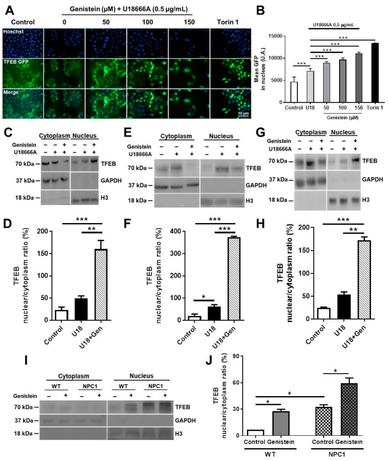
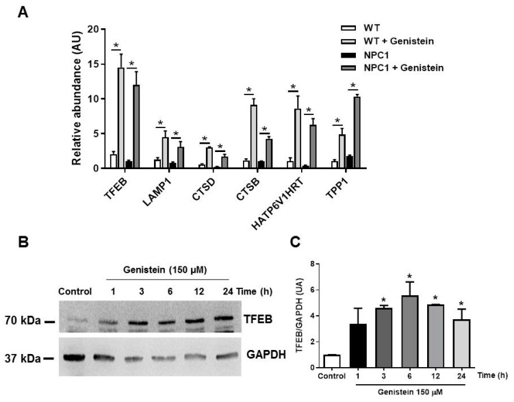
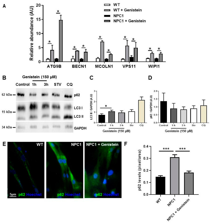
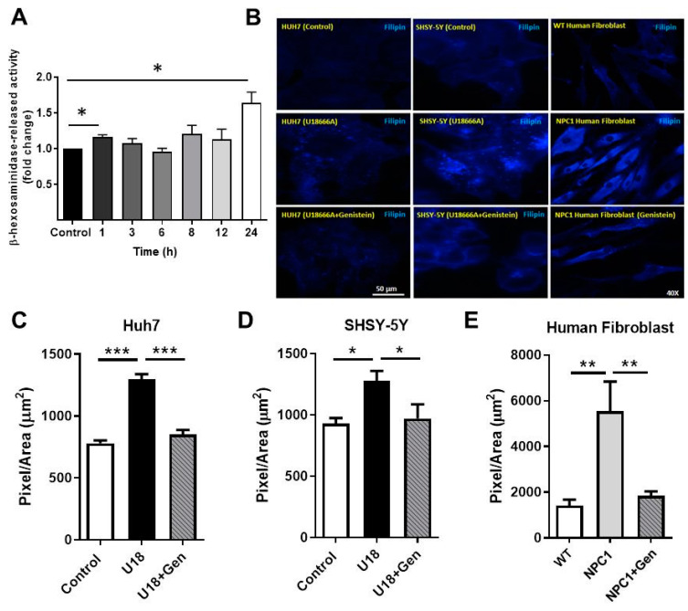
Niemann-Pick type C disease (NPCD) is a lysosomal storage disease (LSD) characterized by abnormal cholesterol accumulation in lysosomes, impaired autophagy flux, and lysosomal dysfunction. The activation of transcription factor EB (TFEB), a master lysosomal function regulator, reduces the accumulation of lysosomal substrates in LSDs where the degradative capacity of the cells is compromised. Genistein can pass the blood-brain barrier and activate TFEB. Hence, we investigated the effect of TFEB activation by genistein toward correcting the NPC phenotype. We show that genistein promotes TFEB translocation to the nucleus in HeLa TFEB-GFP, Huh7, and SHSY-5Y cells treated with U18666A and NPC1 patient fibroblasts. Genistein treatment improved lysosomal protein expression and autophagic flux, decreasing p62 levels and increasing those of the LC3-II in NPC1 patient fibroblasts. Genistein induced an increase in β-hexosaminidase activity in the culture media of NPC1 patient fibroblasts, suggesting an increase in lysosomal exocytosis, which correlated with a decrease in cholesterol accumulation after filipin staining, including cells treated with U18666A and NPC1 patient fibroblasts. These results support that genistein-mediated TFEB activation corrects pathological phenotypes in NPC models and substantiates the need for further studies on this isoflavonoid as a potential therapeutic agent to treat NPCD and other LSDs with neurological compromise.

Keywords: Niemann–Pick C; TFEB; cholesterol; genistein; lysosomal storage diseases; lysosome clearance.


Conflict of interest statement

The authors declare no conflict of interest.

Figures

Figure 1
Figure 1
Genistein promotes transcription factor EB (TFEB) nuclear translocation to the nucleus in Niemann–Pick type C (NPC) cells. Representative images and quantification (A,B) of the TFEB-GFP translocation assay in HeLa cells stably expressing TFEB-GFP treated with 50, 100, and 150 µM of genistein, in control conditions or treated with U18666A (U18) 0.5 µg/mL for 24 h at the same time. As a positive control for TFEB nuclear translocation, we used 0.3 µM Torin 1 for 3 h. TFEB-GFP nuclear localization was analyzed using a high-content nuclear translocation assay in an epifluorescence automated microscope. Scale bar: 10 μm, GFP (green) and Hoechst (blue). For each condition, 3000–5000 cells were analyzed (five wells × 16 images). Representative immunoblot and quantification of endogenous TFEB levels in lysates from nuclear and cytoplasmatic fractions obtained after 24 h treatment with 150 µM of genistein, in control or 0.5 µg/mL of U18 treated HeLa cells stably expressing TFEB-GFP (C,D), Huh7 cells (E,F), SHSY-5Y cells (G,H), GM05659 WT, and the GM03123 NPC1 patient fibroblasts (I,J). Human Glyceraldehyde-3-phosphate dehydrogenase (GAPDH) and Histone H3 were used as cytoplasmatic and nuclear loading controls, respectively. * p ≤ 0.05, ** p ≤ 0.01, *** p ≤ 0.001 data represent mean ± SEM.
Figure 2
Figure 2
Genistein induces the mRNA expression of transcription factor EB (TFEB)-target genes and increases TFEB levels in NPC1 patient fibroblasts. (A) The graph shows qPCR analysis of mRNA levels of different TFEB target genes in GM05659 wild type (WT) fibroblast and GM03123 NPC1 patient fibroblasts after 24 h treatment with 150 µM of genistein. The bar graphs represent the relative fold induction of these genes normalized with GAPDH mRNA levels. Representative immunoblot (B) and quantification (C) of endogenous TFEB total levels in lysates from WT fibroblast and NPC1 patients fibroblast obtained after 1, 3, 6, 12, and 24 h treatment with 150 µM of genistein. GAPDH was used as a loading control. Data were presented as mean ± SEM of three independent samples (each with triplicates), * p ≤ 0.05, analyzed by ANOVA with Tukey posttest, n = 3 for each experiment (in triplicates for genes).
Figure 3
Figure 3
Genistein induces autophagy markers in NPC1 patient fibroblasts. The mRNA levels of the autophagy-related genes, ATG9B, BECN1, MCOLN1, VSP11, and WIPI1, were measured after 24 h of treatment with 150 µM of genistein by qPCR (A). Representative immunoblot (B) and quantification of LC3 (C) and p62 (D) levels in GM05659 wild type (WT) fibroblast and GM03123 NPC1 patient fibroblast obtained after 1 and 3 h treatment with 150 µM of genistein. As a positive control for autophagy inhibition and autophagy activation, we used chloroquine 20 µM and starvation media, respectively, for 3 h. GAPDH was used as loading control. Representative images (E) and quantification (F) of normalized relative fluorescence intensities for p62 on WT and NPC1 patient fibroblasts obtained after 3 h of treatment with 150 µM of genistein. The bar graphs show mean ± SEM, * p ≤ 0.05 and *** p ≤ 0.001 versus control analyzed by ANOVA with Tukey test, n = 3 for each experiment (in triplicates for genes).
Figure 4
Figure 4
TFEB activation by genistein induces lysosomal exocytosis and reduces cholesterol accumulation in NPC models. (A) GM03123 NPC1 patient fibroblasts were cultured in 24-well plates at 30,000 cells/well and treated with 150 µM genistein for 1, 3, 6, 8, 12, and 24 h. The activity of the lysosomal enzyme β-Hexosaminidase was determined in both conditioned media and cell extracts. The bar graphs show the released enzyme activity measured as a percentage of total activity and expressed as fold change. Data are mean ± SEM from independent experiments, each in triplicate. Representative images and quantification of cholesterol accumulation by filipin staining in Huh7 (B,C) and SHSY-5Y (BD) cells in control condition, treated with U18 (0.5 µg/mL) and U18 and genistein and WT (control) and NPC1 patient fibroblasts (BE) treated with genistein 150 µM for 24 h. The filipin intensity quantification was made by the Image J program. The bar graphs show mean ± SEM filipin signal intensity normalized to controls, n = 5 images/condition. * p ≤ 0.05, ** p ≤ 0.01, *** p ≤ 0.001 versus control analyzed by ANOVA with Tukey posttest.

References

    1. Winkler M.B., Kidmose R.T., Szomek M., Thaysen K., Rawson S., Muench S.P., Wüstner D., Pedersen B.P. Structural Insight into Eukaryotic Sterol Transport through Niemann-Pick Type C Proteins. Cell. 2019;179:485–497.e18. doi: 10.1016/j.cell.2019.08.038. - DOI - PubMed
    1. Liao G., Yao Y., Liu J., Yu Z., Cheung S., Xie A., Liang X., Bi X. Cholesterol accumulation is associated with lysosomal dysfunction and autophagic stress in NPC1 -/- mouse brain. Am. J. Pathol. 2007;171:962–975. doi: 10.2353/ajpath.2007.070052. - DOI - PMC - PubMed
    1. Vázquez M.C., Balboa E., Alvarez A.R., Zanlungo S. Oxidative Stress: A Pathogenic Mechanism for Niemann-Pick Type C Disease. Oxidative Med. Cell. Longev. 2012;2012:205713. doi: 10.1155/2012/205713. - DOI - PMC - PubMed
    1. Meske V., Erz J., Priesnitz T., Ohm T.-G. The autophagic defect in Niemann–Pick disease type C neurons differs from somatic cells and reduces neuronal viability. Neurobiol. Dis. 2014;64:88–97. doi: 10.1016/j.nbd.2013.12.018. - DOI - PubMed
    1. Torres S., Balboa E., Zanlungo S., Enrich C., Garcia-Ruiz C., Fernandez-Checa J.C. Lysosomal and Mitochondrial Liaisons in Niemann-Pick Disease. Front. Physiol. 2017;8:982. doi: 10.3389/fphys.2017.00982. - DOI - PMC - PubMed

MeSH terms

Substances

LinkOut - more resources