

Ecklonia cava Extract and Its Derivative Dieckol Promote Vasodilation by Modulating Calcium Signaling and PI3K/AKT/eNOS Pathway in In Vitro and In Vivo Models
- PMID: 33921856
- PMCID: PMC8073412
- DOI: 10.3390/biomedicines9040438
Ecklonia cava Extract and Its Derivative Dieckol Promote Vasodilation by Modulating Calcium Signaling and PI3K/AKT/eNOS Pathway in In Vitro and In Vivo Models
Abstract
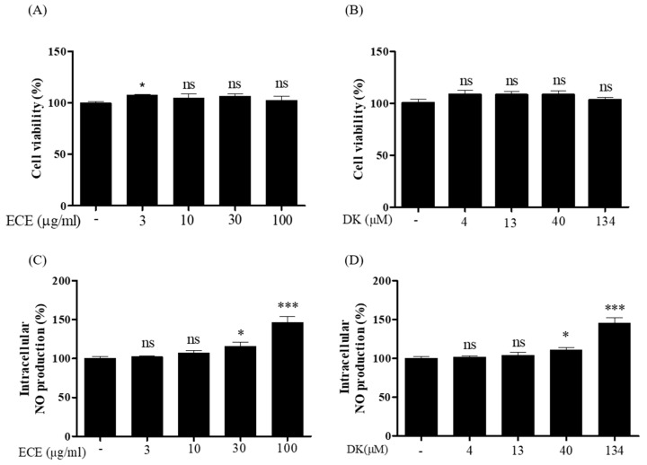
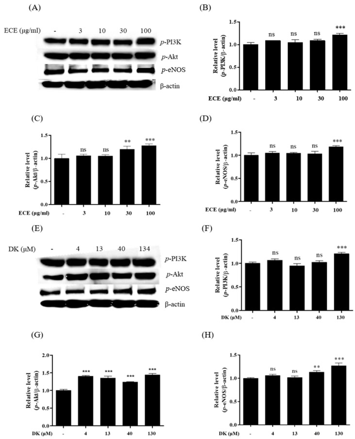
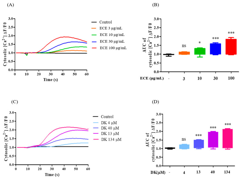
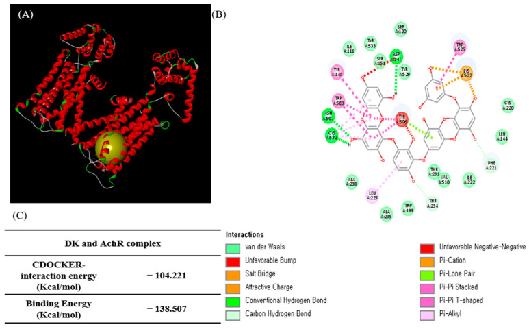
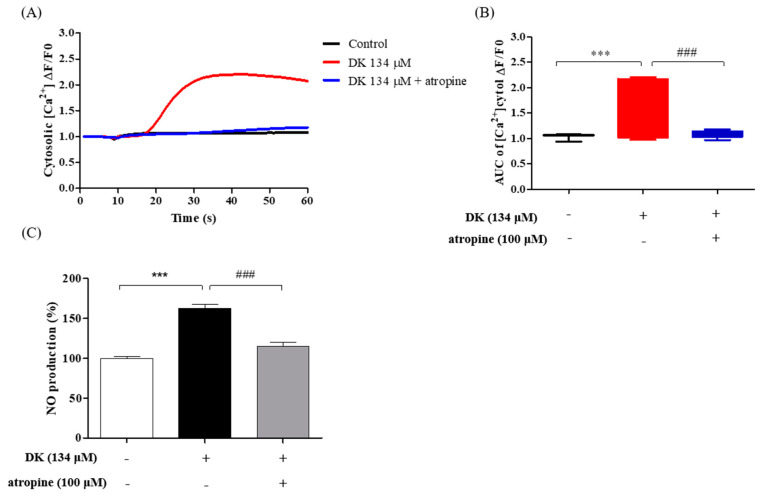
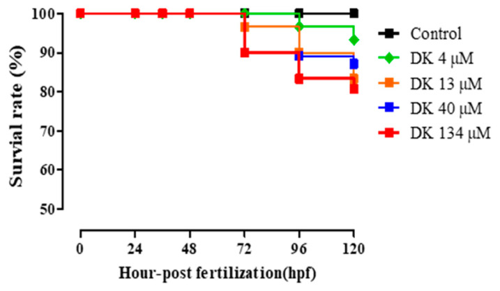
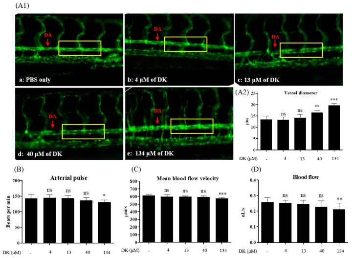
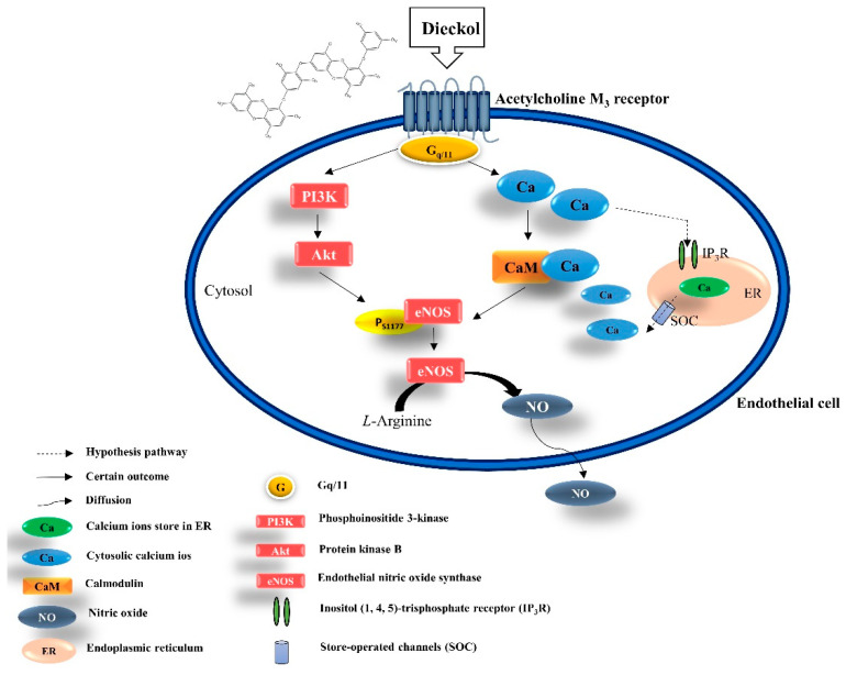
Nitric oxide (NO), an endothelial-derived relaxing factor synthesized by endothelial nitric oxide synthase (eNOS) in endothelial cells, enhances vasodilation by modulating vascular tone. The calcium concentration critically influences eNOS activation in endothelial cells. Thus, modulation of calcium-dependent signaling pathways may be a potential therapeutic strategy to enhance vasodilation. Marine algae reportedly possess protective effects against cardiovascular disorders, including hypertension and vascular dysfunction; however, the underlying molecular signaling pathways remain elusive. In the present study, we extracted and isolated dieckol from Ecklonia cava and investigated calcium transit-enhanced vasodilation. Calcium modulation via the well-known M3 muscarinic acetylcholine receptor (AchM3R), which is linked to NO formation, was investigated and the vasodilatory effect of dieckol was verified. Our results indicated that dieckol effectively promoted NO generation via the PI3K/Akt/eNOS axis and calcium transients influenced by AchM3R. We also treated Tg(flk: EGFP) transgenic zebrafish with dieckol to assess its vasodilatory effect. Dieckol promoted vasodilation by enlarging the dorsal aorta diameter, thus regulating blood flow velocity. In conclusion, our findings suggest that dieckol modulates calcium transit through AchM3R, increases endothelial-dependent NO production, and efficiently enhances vasodilation. Thus, E. cava and its derivative, dieckol, can be considered as potential natural vasodilators.
Keywords: Ecklonia cava; M3 muscarinic acetylcholine receptor; NO production; calcium transit; dieckol; endothelial cell; vasodilation; zebrafish.
Conflict of interest statement
The authors declare no conflict of interest.
Figures
Similar articles
-
Diphlorethohydroxycarmalol Isolated from Ishige okamurae Exerts Vasodilatory Effects via Calcium Signaling and PI3K/Akt/eNOS Pathway.Int J Mol Sci. 2021 Feb 5;22(4):1610. doi: 10.3390/ijms22041610. Int J Mol Sci. 2021. PMID: 33562632 Free PMC article.
-
Protective effect of dieckol isolated from Ecklonia cava against ethanol caused damage in vitro and in zebrafish model.Environ Toxicol Pharmacol. 2013 Nov;36(3):1217-26. doi: 10.1016/j.etap.2013.09.018. Epub 2013 Oct 7. Environ Toxicol Pharmacol. 2013. PMID: 24189014
-
Astragaloside IV Improves Vasodilatation Function by Regulating the PI3K/Akt/eNOS Signaling Pathway in Rat Aorta Endothelial Cells.J Vasc Res. 2018;55(3):169-176. doi: 10.1159/000489958. Epub 2018 Jul 4. J Vasc Res. 2018. PMID: 29972829
-
Marine Alga Ecklonia cava Extract and Dieckol Attenuate Prostaglandin E2 Production in HaCaT Keratinocytes Exposed to Airborne Particulate Matter.Antioxidants (Basel). 2019 Jun 21;8(6):190. doi: 10.3390/antiox8060190. Antioxidants (Basel). 2019. PMID: 31234405 Free PMC article.
-
Anti-hyperlipidemic Effect of Polyphenol Extract (Seapolynol(™)) and Dieckol Isolated from Ecklonia cava in in vivo and in vitro Models.Prev Nutr Food Sci. 2012 Mar;17(1):1-7. doi: 10.3746/pnf.2012.17.1.001. Prev Nutr Food Sci. 2012. PMID: 24471056 Free PMC article.
Cited by
-
Mining Important Herb Combinations of Traditional Chinese Medicine against Hypertension Based on the Symptom-Herb Network Combined with Network Pharmacology.Evid Based Complement Alternat Med. 2022 Mar 22;2022:5850899. doi: 10.1155/2022/5850899. eCollection 2022. Evid Based Complement Alternat Med. 2022. PMID: 35360657 Free PMC article.
-
Sakuranetin as a Potential Regulator of Blood Pressure in Spontaneously Hypertensive Rats by Promoting Vasorelaxation through Calcium Channel Blockade.Biomedicines. 2024 Feb 1;12(2):346. doi: 10.3390/biomedicines12020346. Biomedicines. 2024. PMID: 38397948 Free PMC article.
-
The significance of calcium ions in cerebral ischemia-reperfusion injury: mechanisms and intervention strategies.Front Mol Biosci. 2025 May 12;12:1585758. doi: 10.3389/fmolb.2025.1585758. eCollection 2025. Front Mol Biosci. 2025. PMID: 40421420 Free PMC article. Review.
-
Novel mathematical approach to accurately quantify 3D endothelial cell morphology and vessel geometry based on fluorescently marked endothelial cell contours: Application to the dorsal aorta of wild-type and Endoglin-deficient zebrafish embryos.PLoS Comput Biol. 2024 Aug 30;20(8):e1011924. doi: 10.1371/journal.pcbi.1011924. eCollection 2024 Aug. PLoS Comput Biol. 2024. PMID: 39213451 Free PMC article.
-
Role of medicinal plants in inhibiting SARS-CoV-2 and in the management of post-COVID-19 complications.Phytomedicine. 2022 Apr;98:153930. doi: 10.1016/j.phymed.2022.153930. Epub 2022 Jan 5. Phytomedicine. 2022. PMID: 35114450 Free PMC article.
References
-
- Sauceda A.E.Q., Sáyago-Ayerdi S.G., Ayala-Zavala J.F., Wall-Medrano A., De La Rosa L.A., González-Aguilar G.A., Álvarez-Parrilla E. Biological Actions of Phenolic Compounds. Fruit Veg. Phytochem. 2017;2:125–138. doi: 10.1002/9781119158042.ch6. - DOI
Grants and funding
LinkOut - more resources
Full Text Sources
Other Literature Sources
Molecular Biology Databases
Miscellaneous
