. 2021 Apr 19;9(4):439.
doi: 10.3390/biomedicines9040439.

In Vitro Investigation of Vascular Permeability in Endothelial Cells from Human Artery, Vein and Lung Microvessels at Steady-State and Anaphylactic Conditions

Affiliations

In Vitro Investigation of Vascular Permeability in Endothelial Cells from Human Artery, Vein and Lung Microvessels at Steady-State and Anaphylactic Conditions

Katrine T Callesen et al. Biomedicines. .

Abstract

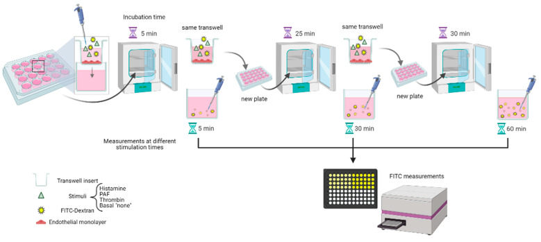
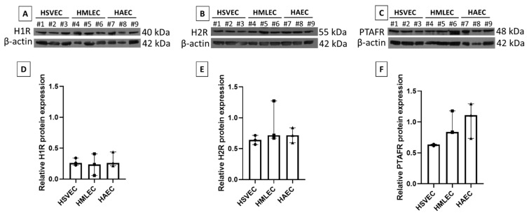
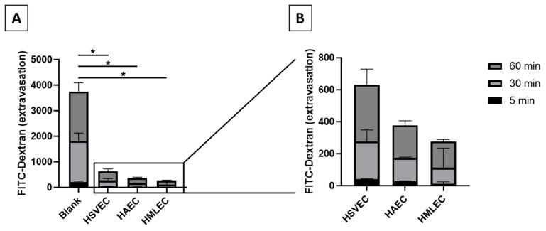
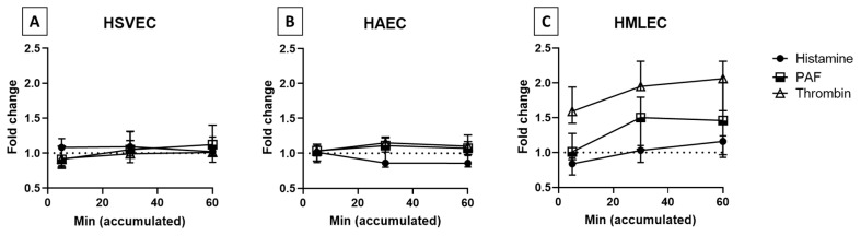
Human anaphylactic reactions largely involve an increase in vascular permeability, which is mainly controlled by endothelial cells (ECs). Due to the acute and serious nature of human anaphylaxis, in vivo studies of blood vessels must be replaced or supplemented with in vitro models. Therefore, we used a macromolecular tracer assay (MMTA) to investigate the EC permeability of three phenotypes of human ECs: artery (HAECs), vein (HSVECs) and microvessels from lung (HMLECs). ECs were stimulated with two fast-acting anaphylactic mediators (histamine and platelet-activating factor (PAF)) and one longer-lasting mediator (thrombin). At steady-state conditions, HSVEC monolayers were the most permeable and HMLEC the least (15.8% and 8.3% after 60 min, respectively). No response was found in ECs from artery or vein to any stimuli. ECs from microvessels reacted to stimulation with thrombin and also demonstrated a tendency of increased permeability for PAF. There was no reaction for histamine. This was not caused by missing receptor expression, as all three EC phenotypes expressed receptors for both PAF and histamine. The scarce response to fast-acting mediators illustrates that the MMTA is not suitable for investigating EC permeability to anaphylactic mediators.

Keywords: PAF; endothelial cells; histamine; macromolecular tracer assay; thrombin; vascular permeability.


Conflict of interest statement

The authors declare no conflict of interest.

Figures

Figure 1
Figure 1
Diagram showing the flow of work for the macromolecular tracer assay. Created with BioRender.com.
Figure 2
Figure 2
Endothelial receptor expression. Human saphenous vein endothelial cells (HSVEC#1–3), human microvascular lung endothelial cells (HMLEC#4–6), and human arterial endothelial cells (HAEC#7–9) were investigated for histamine 1 receptor, H1R (A,D), histamine 2 receptor, H2R (B,E), and platelet activating factor receptor, PTAFR (C,F). (AC) Western blots of specific protein and loading control (β-actin). (DF) Relative protein expression (normalized to β-actin). All were tested in three independent cell cultures (#1–3).
Figure 3
Figure 3
Extravasation of FITC-Dextran in macromolecular tracer assay. (A) The extravasation of FITC-Dextran was measured in transwells without cells (“Blank”) and compared to transwells with monolayers of basal (unstimulated) ECs. (B) Magnification of basal diffusion experiments. Blank, n = 10; Human saphenous vein endothelial cells (HSVEC), n = 5; human arterial endothelial cells (HAEC), n = 4; and human microvascular lung endothelial cells (HMLEC), n = 5. * p < 0.01.
Figure 4
Figure 4
EC permeability upon stimulation with histamine, platelet activator factor (PAF), and thrombin. (A) Human saphenous vein endothelial cells (HSVEC; n = 5 for histamine and PAF, n = 3 for thrombin), (B) human arterial endothelial cells (HAEC; n = 4 for histamine and PAF, n = 3 for thrombin), and (C) human microvascular lung endothelial cells (HMLEC; n = 5 for histamine and PAF, n = 3 for thrombin) were either stimulated with 10 µM histamine, 10 µM PAF, or 0.1 U/mL thrombin, or were unstimulated (basal). Level of FITC was measured from the outer chamber at the indicated accumulated time. Data are depicted as fold change from the basal samples. Dotted line: basal sample indication.

References

    1. Aird W.C. Phenotypic Heterogeneity of the Endothelium. Circ. Res. 2007;100:158–173. doi: 10.1161/01.RES.0000255691.76142.4a. - DOI - PubMed
    1. Uhlig S., Yang Y., Waade J., Wittenberg C., Babendreyer A., Kuebler W.M. Differential Regulation of Lung Endothelial Permeability in Vitro and in Situ. Cell. Physiol. Biochem. 2014;34:1–19. doi: 10.1159/000362980. - DOI - PubMed
    1. Claesson-Welsh L. Vascular permeability—the essentials. Upsala J. Med Sci. 2015;120:135–143. doi: 10.3109/03009734.2015.1064501. - DOI - PMC - PubMed
    1. Egawa G., Nakamizo S., Natsuaki Y., Doi H., Miyachi Y., Kabashima K. Intravital analysis of vascular permeability in mice using two-photon microscopy. Sci. Rep. 2013;3:srep01932. doi: 10.1038/srep01932. - DOI - PMC - PubMed
    1. Wegener J., Seebach J. Experimental tools to monitor the dynamics of endothelial barrier function: A survey of in vitro approaches. Cell and Tissue Research. 2014;355:485–514. doi: 10.1007/s00441-014-1810-3. - DOI - PubMed

LinkOut - more resources