. 2021 Apr 22;22(9):4361.
doi: 10.3390/ijms22094361.

Micro-Current Stimulation Has Potential Effects of Hair Growth-Promotion on Human Hair Follicle-Derived Papilla Cells and Animal Model

Affiliations

Micro-Current Stimulation Has Potential Effects of Hair Growth-Promotion on Human Hair Follicle-Derived Papilla Cells and Animal Model

Donghyun Hwang et al. Int J Mol Sci. .

Abstract

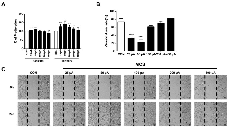
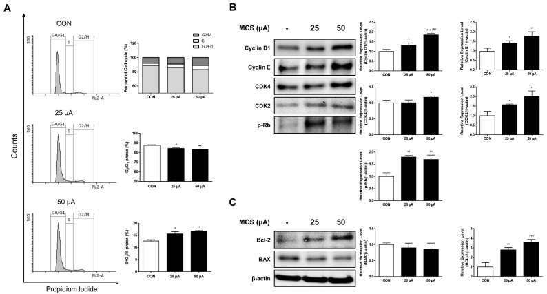
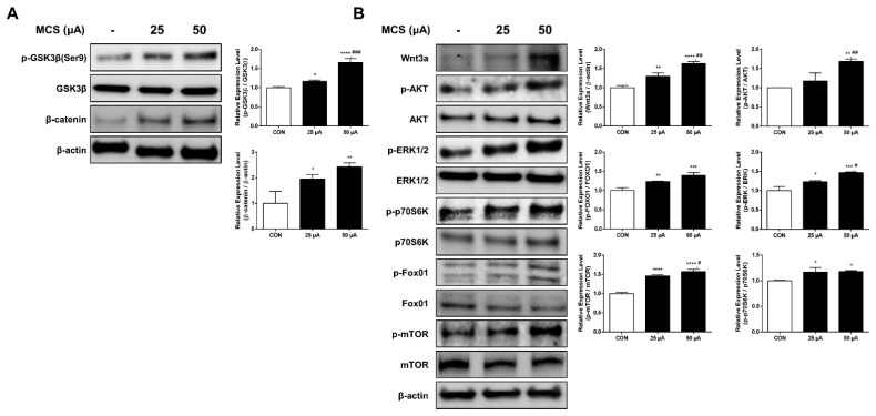
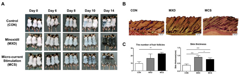
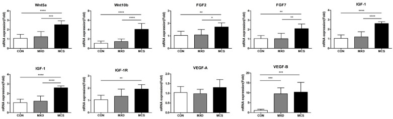
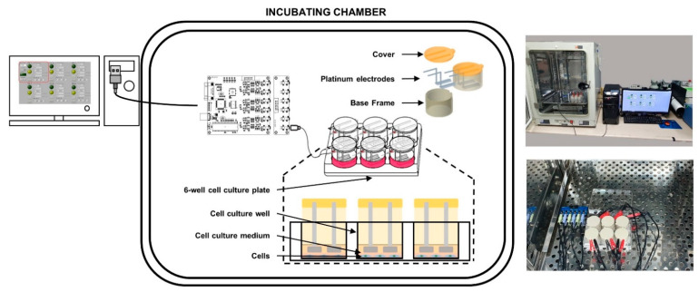
Recently, a variety of safe and effective non-pharmacological methods have been introduced as new treatments of alopecia. Micro-current electrical stimulation (MCS) is one of them. It is generally known to facilitate cell proliferation and differentiation and promote cell migration and ATP synthesis. This study aimed to investigate the hair growth-promoting effect of MCS on human hair follicle-derived papilla cells (HFDPC) and a telogenic mice model. We examined changes in cell proliferation, migration, and cell cycle progression with MCS-applied HFDPC. The changes of expression of the cell cycle regulatory proteins, molecules related to the PI3K/AKT/mTOR/Fox01 pathway and Wnt/β-catenin pathway were also examined by immunoblotting. Subsequently, we evaluated the various growth factors in developing hair follicles by RT-PCR in MCS-applied (MCS) mice model. From the results, the MCS-applied groups with specific levels showed effects on HFDPC proliferation and migration and promoted cell cycle progression and the expression of cell cycle-related proteins. Moreover, these levels significantly activated the Wnt/β-catenin pathway and PI3K/AKT/mTOR/Fox01 pathway. Various growth factors in developing hair follicles, including Wnts, FGFs, IGF-1, and VEGF-B except for VEGF-A, significantly increased in MCS-applied mice. Our results may confirm that MCS has hair growth-promoting effect on HFDPC as well as telogenic mice model, suggesting a potential treatment strategy for alopecia.

Keywords: alopecia; hair growth; human hair follicle dermal papilla cell; micro-current stimulation.


Conflict of interest statement

The authors declare no conflict of interest.

Figures

Figure 1
Figure 1
(A) Effects of MCS on the proliferation of HFDPC cells. To induce cell proliferation, MCS was applied to the HFDPC for 1 h, and the cell was subsequently cultured for 12 and 48 h and incubated further with WST-1 reagent for additional 1 h. The values shown represent the mean ± SD of triplicate measurements of separate experiments. Values are shown as percentages of the control. (B) Relative wound area rate was calculated as the ratio of the remaining wound area at 24 h and the original area at 0 h (C) In vitro scratch assay. Black dotted lines indicate the wound borders at the beginning of the assay and were recorded at 0 and 24 h post-scratching. HFDPC cells were treated with various levels of MCS or left untreated. ** p < 0.01, *** p < 0.001, **** p < 0.0001 vs. control group.
Figure 2
Figure 2
MCS groups promote cell cycle progression and inhibit cell apoptosis. (A) Change in cell cycle distribution in HFDPC following the treatment of MCS. HFDPC were treated with various levels of MCS for 24 h. The analysis of cell cycle distribution was performed by flow cytometry after the staining of DNA by propidium iodide. The population of cells in G0/G1, S, and G2/M phases in MCS-treated HFDPC. * p < 0.05, ** p < 0.001. (B) Immunoblot analysis of cell cycle-related proteins on HFDPC following the treatment of MCS. The cell lysate was analyzed by immunoblot using Cyclin D1, Cyclin E, CDK4, CDK2, and p-Rb antibodies. (C) Immunoblot analysis of cell apoptosis-related proteins on HFDPC following the treatment of MCS. The graph represents the quantitative level of the proteins. * p < 0.05, ** p < 0.01, *** p < 0.001 vs. control group; ## p < 0.01 vs. 25 μA MCS group.
Figure 3
Figure 3
MCS upregulates the GSK3β/β-catenin signaling pathway and PI3K/AKT/mTOR/Fox01 signaling pathway. (A) Immunoblot analysis of the expression of p-GSK3β (Ser9), GSK3β, and β-catenin in HFDPC cells following treatment with MCS. (B) Immunoblot analysis of the expression of Wnt3a, p-AKT, AKT, p-ERK1/2, ERK1/2, p-p70S6K, p70S6K, p-Fox01, Fox01, p-mTOR, and mTOR in HFDPC cells following treatment with MCS. * p < 0.05, ** p < 0.01, *** p < 0.001, **** p < 0.0001 vs. control group; # p < 0.05, ## p < 0.01 vs. 25 μA MCS group.
Figure 4
Figure 4
MCS accelerates the onset of black pigmentation and increases the number of follicles and skin thickness. (A) Photographs of shaved dorsal skin were taken at Day 0, 6, 8, 10 and 14 in control group (CON), minoxidil-treated group (MXD) used as a positive control, and micro-current stimulation with the intensity of 50 μA group (MCS). (B) Longitudinal sections of the dorsal skins for each group by H&E staining. (C) The number of hair follicles and skin thickness (Full-thickness) in the section for each group. * p < 0.05, ** p < 0.01, *** p < 0.001.
Figure 5
Figure 5
MCS increases the mRNA expression levels of growth factors contributing to telogen–anagen transition and hair growth promotion in telogenic mice. The mRNA expression levels were measured in the control group (CON), the minoxidil-treated group (MXD) used as a positive control, and micro-current stimulation with the intensity of the 50 μA group (MCS). All mRNA expression levels were normalized to that of GAPDH mRNA expression and expressed as fold changes relative to that of CON. * p < 0.05, ** p < 0.01, *** p < 0.001, **** p < 0.0001.
Figure 6
Figure 6
MCS system available in incubating chamber.

References

    1. Lei M., Chuong C.-M. Aging, alopecia, and stem cells. Science. 2016;351:559–560. doi: 10.1126/science.aaf1635. - DOI - PubMed
    1. Matsumura H., Mohri Y., Binh N.T., Morinaga H., Fukuda M., Ito M., Nishimura E.K. Hair follicle aging is driven by transepidermal elimination of stem cells via COL17A1 proteolysis. Science. 2016;351:6273. doi: 10.1126/science.aad4395. - DOI - PubMed
    1. Price V.H. Treatment of Hair Loss. N. Engl. J. Med. 1999;341:964–973. doi: 10.1056/NEJM199909233411307. - DOI - PubMed
    1. Mounsey A.L., Reed S.W. Diagnosing and treating hair loss. Am. Fam. Physician. 2009;80:356–362. - PubMed
    1. Sasson M., Shupack J.L., Stiller M.J. status of medical treatment for androgenetic alopecia. Int. J. Dermatol. 1993;32:701–706. doi: 10.1111/j.1365-4362.1993.tb02735.x. - DOI - PubMed

LinkOut - more resources