Review
. 2021 Apr 22;11(5):1083.
doi: 10.3390/nano11051083.

Graphene Oxide: Opportunities and Challenges in Biomedicine

Affiliations
Review

Graphene Oxide: Opportunities and Challenges in Biomedicine

Pariya Zare et al. Nanomaterials (Basel). .

Abstract

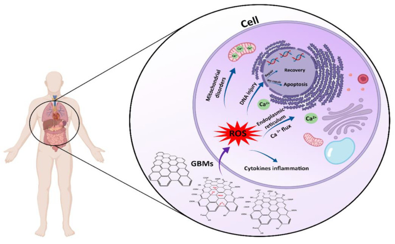
Desirable carbon allotropes such as graphene oxide (GO) have entered the field with several biomedical applications, owing to their exceptional physicochemical and biological features, including extreme strength, found to be 200 times stronger than steel; remarkable light weight; large surface-to-volume ratio; chemical stability; unparalleled thermal and electrical conductivity; and enhanced cell adhesion, proliferation, and differentiation properties. The presence of functional groups on graphene oxide (GO) enhances further interactions with other molecules. Therefore, recent studies have focused on GO-based materials (GOBMs) rather than graphene. The aim of this research was to highlight the physicochemical and biological properties of GOBMs, especially their significance to biomedical applications. The latest studies of GOBMs in biomedical applications are critically reviewed, and in vitro and preclinical studies are assessed. Furthermore, the challenges likely to be faced and prospective future potential are addressed. GOBMs, a high potential emerging material, will dominate the materials of choice in the repair and development of human organs and medical devices. There is already great interest among academics as well as in pharmaceutical and biomedical industries.

Keywords: 3D scaffold; carbon; cell adhesion; functionalization; graphene; graphene oxide; human organs; interface; stem cells.


Conflict of interest statement

The authors declare no conflict of interest.

Figures

Figure 1
Figure 1
Schematic of graphene oxide nanomaterials and their application in tissue engineering, particularly in nerve, muscle, heart, skin, cartilage, dental, and other tissues.
Figure 2
Figure 2
Graphene, graphene oxide, and reduced graphene oxide synthesis methods [9].
Figure 3
Figure 3
Schematic illustration of (A) the LBLC graphene nerve conduit. (a) The green layers are PDA/RGD adhesive macromolecules. The purple layer is single or multilayered graphene and the PCL-blended layer. The blue layer is the graphene and PCL-blended layer once more. (b) The GO/PCL nerve conduit in a sciatic nerve defect model in SD rats. Reused with permission from [30]. Copyright © 2021, Springer Nature. (B) rGO/alginate microgel embedding MSCs for cardiac tissue repair post-MI. Due to the loaded rGO, it was expected that encapsulated MSCs were protected from the severe oxidative stress in the infarcted tissue and facilitated cardiac regeneration. Reused with permission from [27]. Copyright © 2021, Elsevier Ltd. (C) CS-DA-GO composite hydrogel fabrication in three steps, which causes the enhanced properties of the hydrogel. (A) Self-healing mechanism of the hydrogel. (B) Self-adhesiveness of the hydrogel. (C) Enhanced conductivity. Reused with permission from [29]. Copyright © 2021, Elsevier Ltd.
Figure 4
Figure 4
(A) Pseudocolored SEM images of human osteoblasts that successfully adhered to the surface of scaffolds at an upward trend (Days 1, 3, and 7). Reused with permission from [58]. Copyright © 2021, Elsevier Ltd. (B) Surgical procedure of the preparation of the surgical size defect (5 mm) critical in the rat calvarium (ac), placement of the chitosan–graphene oxide scaffold (d), and closure of the periosteum and skin (e,f). Reused with permission from [64]. Copyright © 2021, Springer Nature.
Figure 5
Figure 5
(A) Wound healing of the skin through the treatment of various scaffolds. (a) Model of wound regeneration. Digital images (b) and closure rate (c) of the wound defects; multiple treatments at Days 0, 7, 14, and 21. (d) Depictive images of H&E-stained histological sections after 21 days (arrows indicate the granulation tissue). Reused with permission from [68]. Copyright © 2021 American Chemical Society. (B) (a) A normal rat knee joint; (b) knee joint restoration in different experimental groups (M: without implant; GO-NGO (U): non-crosslinked hydrogel; GO-NGO (T): microplasma crosslinked hydrogel) i 4 and 8 weeks. Reused with permission from [24]. Copyright © 2021 American Chemical Society.
Figure 6
Figure 6
The prohibition effect of GO on biofilm formation. Reused with permission from [90]. Copyright © 2021 WILEY-VCH Verlag GmbH and Co. KGaA, Weinheim.
Figure 7
Figure 7
Graphical conclusion.
Figure 8
Figure 8
Schematic illustration of the cellular toxicity of GOBMs when exposed to the cell. GOBMs enter the cell through various pathways, which affects their shape, size, and surface chemistry and eventually results in ROS production. An increased ROS level may cause mitochondrial membrane depolarization and Ca2+ release; lipid, protein, and DNA damage; and inflammation response by releasing cytokines and chemokines.

References

    1. Dag Line P. The Fundamental Challenges in Organ Transplantation. OBM Transpl. 2017;1:6. doi: 10.21926/obm.transplant.1704006. - DOI
    1. Aleemardani M., Bagher Z., Farhadi M., Chahsetareh H., Najafi R., Eftekhari B., Seifalian A.M. Can tissue engineering bring hope in the development of human tympanic membrane? Tissue Eng. Part B Rev. 2020:1–62. doi: 10.1089/ten.teb.2020.0176. - DOI - PubMed
    1. Peng Z., Zhao T., Zhou Y., Li S., Li J., Leblanc R.M. Bone Tissue Engineering via Carbon-Based Nanomaterials. Adv. Healthc. Mater. 2020;9:1901495. doi: 10.1002/adhm.201901495. - DOI - PubMed
    1. Aydin T., Gurcan C., Taheri H., Yilmazer A. Advances in Experimental Medicine and Biology. Volume 1107. Springer; New York, NY, USA: 2018. Graphene based materials in neural tissue regeneration; pp. 129–142. - PubMed
    1. Bellet P., Gasparotto M., Pressi S., Fortunato A., Scapin G., Mba M., Menna E., Filippini F. Graphene-based scaffolds for regenerative medicine. Nanomaterials. 2021;11:404. doi: 10.3390/nano11020404. - DOI - PMC - PubMed