Review
. 2021 Apr 22;10(5):982.
doi: 10.3390/cells10050982.

Macrophages and Immune Responses in Uterine Fibroids

Affiliations
Review

Macrophages and Immune Responses in Uterine Fibroids

Alessandro Zannotti et al. Cells. .

Abstract

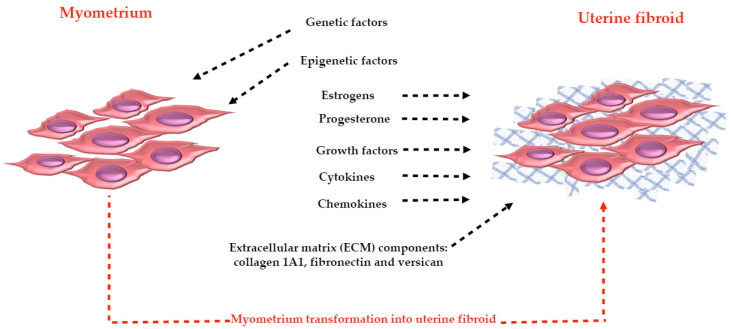
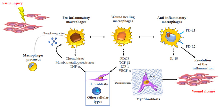
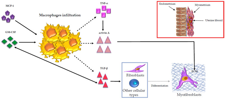
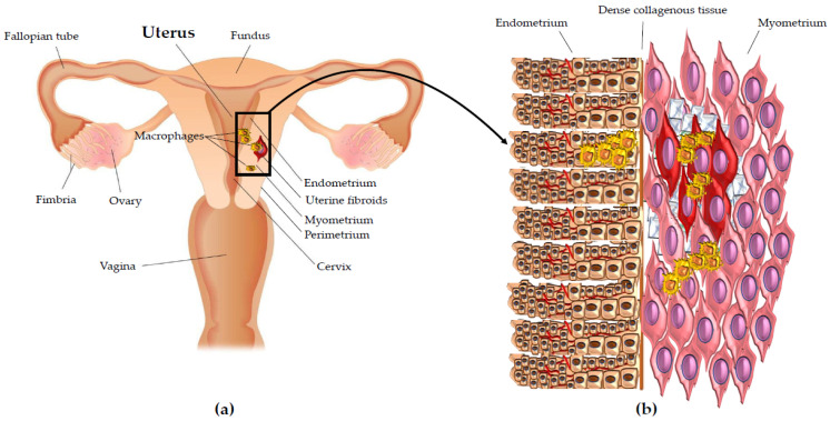
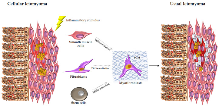
Uterine fibroids represent the most common benign tumors of the uterus. They are considered a typical fibrotic disorder. In fact, the extracellular matrix (ECM) proteins-above all, collagen 1A1, fibronectin and versican-are upregulated in this pathology. The uterine fibroids etiology has not yet been clarified, and this represents an important matter about their resolution. A model has been proposed according to which the formation of an altered ECM could be the result of an excessive wound healing, in turn driven by a dysregulated inflammation process. A lot of molecules act in the complex inflammatory response. Macrophages have a great flexibility since they can assume different phenotypes leading to the tissue repair process. The dysregulation of macrophage proliferation, accumulation and infiltration could lead to an uncontrolled tissue repair and to the consequent pathological fibrosis. In addition, molecules such as monocyte chemoattractant protein-1 (MCP-1), granulocyte macrophage-colony-stimulating factor (GM-CSF), transforming growth factor-beta (TGF-β), activin A and tumor necrosis factor-alfa (TNF-α) were demonstrated to play an important role in the macrophage action within the uncontrolled tissue repair that contributes to the pathological fibrosis that represents a typical feature of the uterine fibroids.

Keywords: ECM; inflammatory process; macrophages; pathological fibrosis; tissue repair; uterine fibroids.


Conflict of interest statement

The authors declare no conflict of interest.

Figures

Figure 1
Figure 1
Illustration of the promoters of fibroid growth. The blue net represents the typical extracellular matrix (ECM) proteins: collagen 1A1, fibronectin and versican. The abundant ECM in uterine fibroids (approximately 50% more than the corresponding myometrium) was suggested to represent a reservoir for the other promoters of fibroid growth.
Figure 2
Figure 2
Illustration of the role of the macrophages and their highly flexible programming in tissue repair and fibrosis in several organs. Macrophages, because of their high flexibility, can play a key regulatory role in every stage that characterizes the tissue repair and fibrosis from the promotion to the resolution of the inflammation leading to the wound closure. The figure shows the principal events and principal molecules: chemokines, Matrix metalloproteinases, tumor necrosis factor-alfa (TNF-α), platelet-derived growth factor (PDGF), transforming growth factor-beta 1 (TGF-β1), insulin-like growth factor-1 (IGF-1), vascular endothelial growth factor-alfa (VEGF-α), programmed death-ligand 1 (PD-L1) and programmed death-ligand 2 (PD-L2), interleukin-10 (IL-10) involved in the process, highlighting the different phenotypic states that the macrophages can assume in the process. The blue net represents the extracellular matrix (ECM) that is produced by myofibroblasts after that fibroblasts or other cellular types differentiated into them.
Figure 3
Figure 3
Illustration of the macrophages’ (yellow in the figure) role in uterine fibroids. Monocyte chemoattractant protein-1 (MCP-1) takes part in the regulation of the macrophages’ infiltration. The granulocyte macrophage-colony-stimulating factor (GM-CSF) is considered the most important growth factor for macrophage proliferation. GM-CSF can establish regulatory interactions with the transforming growth factor-beta (TGF-β), which was shown to be the most important growth factor secreted by macrophages. In uterine fibroids, TGF-β is overexpressed and it contributes to myofibroblast differentiation. Macrophages also secrete activin A, an immuno-regulator belonging to the TGF-β family. Activin A develops a pro-fibrotic action leading to the expression of the extracellular matrix (ECM) proteins (represented by the blue net in the figure), which are overexpressed in uterine fibroids. Activin A mRNA expression in uterine fibroids is upregulated by the tumor necrosis factor-alfa (TNF-α), an inflammatory mediator mainly produced by macrophages. On the right, above the image that portrays the myofibroblasts, the uterine fibroids (red in the figure) with the myometrium (pink in the figure), the endometrium (brown in the figure), the macrophages (yellow in the figure) and the overexpressed ECM proteins (blue net in the figure) are represented. The blood vessels within endometrium are also represented (red lines in the figure).
Figure 4
Figure 4
Macrophages in uterine fibroids. (a) Illustration of uterus showing the macrophage density in uterine fibroids pathology. (b) Enlargement of the detail showing the macrophage density in uterine fibroids pathology. Macrophages (yellow in the figure) predominantly localize inside uterine fibroids (red in the figure) and in the myometrium tissue (pink in the figure) next to them. Autologous distant myometrium shows low levels of macrophage infiltration. The macrophage density is higher also in the endometrium (brown in the figure) next to uterine fibroids than in the autologous endometrium far from uterine fibroid nodules. The extracellular matrix (ECM) around and within the uterine fibroids is also represented (blue net in the figure). The blood vessels within endometrium are also represented (red lines in the figure).
Figure 5
Figure 5
Illustration of the possible phase mechanism of leiomyoma development proposed by our group. Cellular leiomyoma is considered as the first step in the tumoral transformation. In fact, cellular leiomyoma shows higher levels of macrophage (yellow in the figure) infiltration and an increased number of inflammatory cells. This aspect could represent a response to an inflammatory stimulus that leads some cellular leiomyoma cells to myofibroblast differentiation with the consequent upregulation of the typical extracellular matrix (ECM) proteins. In fact, usual leiomyoma shows a larger amount of ECM proteins and low levels of macrophage infiltration. So, usual leiomyoma could be considered as the late-phase tumor. The blue net represents the typical ECM proteins: collagen 1A1, fibronectin and versican. The red color represents the uterine fibroids (light red for cellular leiomyoma histotype and dark red for usual leiomyoma histotype). The pink color represents the myometrium; the brown color represents the endometrium. The blood vessels within endometrium are also represented (red lines in the figure).

References

    1. McEvoy A., Sabir S. Physiology, Pregnancy Contractions. StatPearls; Treasure Island, FL, USA: 2021. - PubMed
    1. Valente R., Malesani M.G. Dizionario Medico. Larousse; Paris, France: 1984. p. 928.
    1. Gentile F. Enciclopedia Italiana. Volume 16. Grolier; New Delhi, India: 1987. p. 271.
    1. Day Baird D., Dunson D.B., Hill M.C., Cousins D., Schectman J.M. High cumulative incidence of uterine leiomyoma in black and white women: Ultrasound evidence. Am. J. Obstet. Gynecol. 2003;188:100–107. doi: 10.1067/mob.2003.99. - DOI - PubMed
    1. Stewart E.A. Uterine fibroids. Lancet. 2001;357:293–298. doi: 10.1016/S0140-6736(00)03622-9. - DOI - PubMed