. 2021 Apr 22;9(5):459.
doi: 10.3390/biomedicines9050459.

Ferulic Acid Induces Keratin 6α via Inhibition of Nuclear β-Catenin Accumulation and Activation of Nrf2 in Wound-Induced Inflammation

Affiliations

Ferulic Acid Induces Keratin 6α via Inhibition of Nuclear β-Catenin Accumulation and Activation of Nrf2 in Wound-Induced Inflammation

Kang-Hoon Kim et al. Biomedicines. .

Abstract

Injured tissue triggers complex interactions through biological process associated with keratins. Rapid recovery is most important for protection against secondary infection and inflammatory pain. For rapid wound healing with minimal pain and side effects, shilajit has been used as an ayurvedic medicine. However, the mechanisms of rapid wound closure are unknown. Here, we found that shilajit induced wound closure in an acute wound model and induced migration in skin explant cultures through evaluation of transcriptomics via microarray testing. In addition, ferulic acid (FA), as a bioactive compound, induced migration via modulation of keratin 6α (K6α) and inhibition of β-catenin in primary keratinocytes of skin explant culture and injured full-thickness skin, because accumulation of β-catenin into the nucleus acts as a negative regulator and disturbs migration in human epidermal keratinocytes. Furthermore, FA alleviated wound-induced inflammation via activation of nuclear factor erythroid-2-related factor 2 (Nrf2) at the wound edge. These findings show that FA is a novel therapeutic agent for wound healing that acts via inhibition of β-catenin in keratinocytes and by activation of Nrf2 in wound-induced inflammation.

Keywords: K6α; Nrf2; ferulic acid; keratinocytes; shilajit; wound healing; β-catenin.


Conflict of interest statement

The authors declare no conflict of interest.

Figures
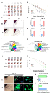
Figure 1
Figure 1
Wound healing of SH and FA. (a) Representative digital images of acute wounds treated with SH on days 0, 1, 3, 5, and 7 post-wounding. (b) Closure of acute wounds with SH was presented as percentage of wound area from the initial wound size. Mean ± SEM; n = 8; *, p < 0.05; **, p < 0.01; ***, p < 0.001 compared with basal group. Statistical analysis was performed using two-way ANOVA, Bonferroni’s test, or paired t-test. (c) Representative data of mouse skin explants processed for hematoxylin and eosin (H&E) staining at day 12 and day 24 in skin explant culture. The skin explant culture was analyzed in triplicate. (d) The quantitation of cellular outgrowth from explants was enhanced with SH. (e) The cells derived from skin explants were analyzed for Krt6α using qPCR at day 12 and day 24. Mean ± SEM; n = 6; *, p < 0.05. The significance of the outgrowth area was calculated using ImageJ software and analyzed using a paired t-test. (f,g) The classification criteria for the 17 categories were provided in the GO ontology databases (http://www.geneontology.org). (h) Representative digital images of acute wounds treated with SH on days 0, 1, 3, 5, and 7 post-wounding. (i) Closure of acute wounds with FA was presented as percentage of wound area from the initial wound size. Mean ± SEM; n = 8; *, p < 0.05; **, p < 0.01; ***, p < 0.001 compared with basal group. Statistical analysis was performed using two-way ANOVA, Bonferroni’s test, or paired t-test. BSL: basal, Ve: vehicle (poly petroleum jelly), FA: ferulic acid,%: (w/w). (j) Examples of mouse skin explants processed for hematoxylin and eosin (H&E) staining and immunofluorescence analyses of K6β after day 12 in ex vivo skin explant culture. (k) The quantitation of cell outgrowth from explants was enhanced with FA. The skin explant culture was analyzed in triplicate. Mean ± SEM; n = 6; ***, p < 0.001. The significance of outgrowth was calculated using ImageJ software and analyzed using paired t-test. (l) The cells derived from the ex vivo skin explant culture were analyzed for β-catenin using qPCR at day 12. Mean ± SEM; n = 6; ***, p < 0.001. The significance was analyzed using paired t-test.
Figure 2
Figure 2
SH and FA induce epithelialization of keratinocytes. (a,b) Immunofluorescence (IF) staining of K6α and β-catenin in the epithelium at day 3 or 7 after wounding with or without SH. (c,d) Protein expression of K6α and β-catenin was analyzed after exposure to SH using Western blotting at day 3 and day 7. (e,f) K6α and β-catenin in the wounded epithelium at day 3 or 7 after exposure to FA. (g,h) Protein expression of K6α and β-catenin was analyzed after exposure to FA using Western blotting at day 3 and day 7. The IF and Western blots were analyzed in triplicate. Mean ± SEM; n = 5; epi: epidermis; derm: dermis.
Figure 3
Figure 3
FA suppresses nucleus translocation of β-catenin in primary keratinocytes but induces nucleus translocation of β-catenin in primary fibroblasts. (a) After treatment with or without LiCl, immunofluorescence (IF) staining of c-myc and β-catenin was examined in primary keratinocytes. (b) The graph indicates the nucleus translocation ratio (%) of β-catenin. (c) The graph indicates the nucleus translocation ratio (%) of c-myc. Significance was measured using t-tests. Note: ** p < 0.01, scale bar: 10μm. The IF was analyzed in triplicate. Mean ± SEM; n = 100. (d) Protein expression of c-myc or β-catenin was analyzed using Western blotting in primary keratinocytes. Significance was measured using t-tests. Note: ** p<0.01. (e,f) Protein expression of β-catenin and c-myc was analyzed using Western blotting in primary keratinocytes. The Western blots were analyzed in triplicate. Mean ± SEM; n = 5. Significance was measured using t-tests. Note: * p < 0.05, ** p < 0.01. (g) After treatment with or without LiCl, immunofluorescence (IF) staining of c-myc and β-catenin was examined in primary fibroblasts. (h) The graph indicates the nucleus translocation ratio (%) of β-catenin in primary fibroblasts. (i) The graph indicates the nucleus translocation ratio (%) of c-myc in primary fibroblasts. Significance was measured using t-tests. Note: ** p < 0.01, scale bar: 30μm. The IF was analyzed in triplicate. Mean ± SEM; n = 100. (j) Protein expression of c-myc or β-catenin was analyzed using Western blotting in primary fibroblasts. (k,l) Protein expression of β-catenin and c-myc was analyzed using Western blotting in primary fibroblasts. The IF and Western blot samples were analyzed in triplicate. Mean ± SEM; n = 5. Significance was measured using t-tests. Note: * p < 0.05.
Figure 4
Figure 4
FA has anti-inflammatory effects via activation of NRF2. (a) Immunofluorescence (IF) staining of COX-2 and iNOS indicated in the epithelium at day 3 or 7 after wounding with or without FA. (b) Immunofluorescence (IF) staining of Nrf2 indicated in the epithelium at day 3 or 7 after wounding with or without FA. (c) Protein expression of COX-2 and iNOS was analyzed after exposure to FA using Western blotting in RAW 264.7 cells. (d) K6α and β-catenin are indicated in the wounded epithelium at day 3 or 7 after exposure to FA. COX-2 and iNOS expression were observed using Western blotting under Nfe2l2 knockdown conditions. (e) The graph showed the knockdown ratio of Nfe2l2. The IF, Western blotand Nfe2l2 knockdown test were performed in triplicate. Mean ± SEM; ** p < 0.01, n = 5. epi: epidermis, derm: dermis.
Figure 5
Figure 5
SH and FA enhance cellular proliferation. (a) The curves represent the course of the murine keratinocytes’ (kera 308 cell line) viability with SH for 72 h. (b) FA’s effects were assessed for 72 h. The RTCA was analyzed in triplicate. Each significance was measured using one-way ANOVA; n = 3, *** p < 0.001, ** p < 0.01, C/W; cells per well, arrow, treated point; BSL, basal; SH, shilajit; FA, ferulic acid. The graphs treated with SH or FA were compared with the area under curve using ImageJ software. (c) Immunofluorescence (IF) staining of Ki67 indicated in the wound epithelium at day 7. Significance was measured using t-tests. The IF was analyzed in triplicate. Mean ± SEM; n = 5, * p< 0.05, ** p < 0.01.
Figure 5
Figure 5
SH and FA enhance cellular proliferation. (a) The curves represent the course of the murine keratinocytes’ (kera 308 cell line) viability with SH for 72 h. (b) FA’s effects were assessed for 72 h. The RTCA was analyzed in triplicate. Each significance was measured using one-way ANOVA; n = 3, *** p < 0.001, ** p < 0.01, C/W; cells per well, arrow, treated point; BSL, basal; SH, shilajit; FA, ferulic acid. The graphs treated with SH or FA were compared with the area under curve using ImageJ software. (c) Immunofluorescence (IF) staining of Ki67 indicated in the wound epithelium at day 7. Significance was measured using t-tests. The IF was analyzed in triplicate. Mean ± SEM; n = 5, * p< 0.05, ** p < 0.01.

Similar articles

Cited by

References

    1. Demidova-Rice T.N., Hamblin M.R., Herman I.M. Acute and Impaired Wound Healing: Pathophysiology and current methods for drug delivery, part 1: Normal and chronic wounds: Biology, causes, and approaches to care. Adv. Ski. Wound Care. 2012;25:304–314. doi: 10.1097/01.ASW.0000416006.55218.d0. - DOI - PMC - PubMed
    1. Elliot S., Wikramanayake T.C., Jozic I., Tomic-Canic M. A Modeling Conundrum: Murine Models for Cutaneous Wound Healing. J. Investig. Dermatol. 2018;138:736–740. doi: 10.1016/j.jid.2017.12.001. - DOI - PubMed
    1. Paladini R.D., Takahashi K., Bravo N.S., Coulombe P. Onset of re-epithelialization after skin injury correlates with a reorganization of keratin filaments in wound edge keratinocytes: Defining a potential role for keratin 16. J. Cell Biol. 1996;132:381–397. doi: 10.1083/jcb.132.3.381. - DOI - PMC - PubMed
    1. Takahashi K., Yan B., Yamanishi K., Imamura S., Coulombe P.A. The Two Functional Keratin 6 Genes of Mouse Are Differentially Regulated and Evolved Independently from Their Human Orthologs. Genomics. 1998;53:170–183. doi: 10.1006/geno.1998.5476. - DOI - PubMed
    1. Wong P., Coulombe P.A. Loss of keratin 6 (K6) proteins reveals a function for intermediate filaments during wound repair. J. Cell Biol. 2003;163:327–337. doi: 10.1083/jcb.200305032. - DOI - PMC - PubMed

LinkOut - more resources