

Senescent Microglia: The Key to the Ageing Brain?
- PMID: 33922383
- PMCID: PMC8122783
- DOI: 10.3390/ijms22094402
Senescent Microglia: The Key to the Ageing Brain?
Abstract
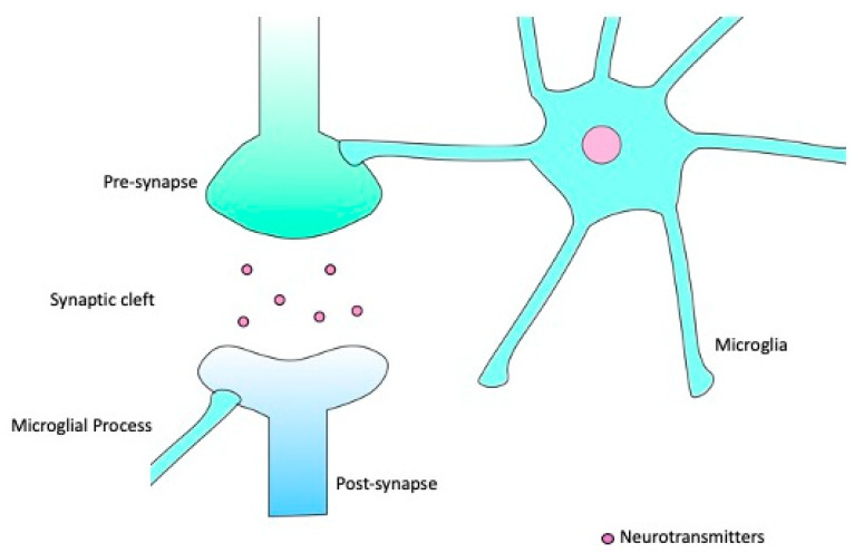
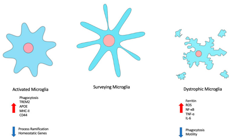
Ageing represents the single biggest risk factor for development of neurodegenerative disease. Despite being such long-lived cells, microglia have been relatively understudied for their role in the ageing process. Reliably identifying aged microglia has proven challenging, not least due to the diversity of cell populations, and the limitations of available models, further complicated by differences between human and rodent cells. Consequently, the literature contains multiple descriptions and categorisations of microglia with neurotoxic phenotypes, including senescence, without any unifying markers. The role of microglia in brain homeostasis, particularly iron storage and metabolism, may provide a key to reliable identification.
Keywords: Senescence Associated Secretory Phenotype; ageing; iron; microglia; neurodegeneration; senescence.
Conflict of interest statement
The authors declare no conflict of interest.
Figures
References
-
- Priller J., Flügel A., Wehner T., Boentert M., Haas C.A., Prinz M., Fernández-Klett F., Prass K., Bechmann I., De Boer B.A., et al. Targeting gene-modified hematopoietic cells to the central nervous system: Use of green fluorescent protein uncovers microglial engraftment. Nat. Med. 2001;7:1356–1361. doi: 10.1038/nm1201-1356. - DOI - PubMed
-
- McKercher S.R., Torbett B.E., Anderson K.L., Henkel G.W., Vestal D.J., Baribault H., Klemsz M., Feeney A.J., Wu G.E., Paige C.J., et al. Targeted disruption of the PU.1 gene results in multiple hematopoietic abnormalities. EMBO J. 1996;15:5647–5658. doi: 10.1002/j.1460-2075.1996.tb00949.x. - DOI - PMC - PubMed
Publication types
MeSH terms
LinkOut - more resources
Full Text Sources
Medical
