. 2021 Apr 22;7(5):325.
doi: 10.3390/jof7050325.

Characterization of a Dye-Decolorizing Peroxidase from Irpex lacteus Expressed in Escherichia coli: An Enzyme with Wide Substrate Specificity Able to Transform Lignosulfonates

Affiliations

Characterization of a Dye-Decolorizing Peroxidase from Irpex lacteus Expressed in Escherichia coli: An Enzyme with Wide Substrate Specificity Able to Transform Lignosulfonates

Laura Isabel de Eugenio et al. J Fungi (Basel). .

Abstract

A dye-decolorizing peroxidase (DyP) from Irpex lacteus was cloned and heterologously expressed as inclusion bodies in Escherichia coli. The protein was purified in one chromatographic step after its in vitro activation. It was active on ABTS, 2,6-dimethoxyphenol (DMP), and anthraquinoid and azo dyes as reported for other fungal DyPs, but it was also able to oxidize Mn2+ (as manganese peroxidases and versatile peroxidases) and veratryl alcohol (VA) (as lignin peroxidases and versatile peroxidases). This corroborated that I. lacteus DyPs are the only enzymes able to oxidize high redox potential dyes, VA and Mn+2. Phylogenetic analysis grouped this enzyme with other type D-DyPs from basidiomycetes. In addition to its interest for dye decolorization, the results of the transformation of softwood and hardwood lignosulfonates suggest a putative biological role of this enzyme in the degradation of phenolic lignin.

Keywords: DyP; fungi; lignin; lignocellulosic biomass; oxidoreductases.


Conflict of interest statement

The authors declare no conflict of interest.

Figures

Figure 1
Figure 1
Catalytic cycle of peroxidases adapted from [9]. RS: resting state, CI: compound I; CII, compound II.
Figure 2
Figure 2
Sequence alignment including DyPs from I. lacteus (IrlacDyP), A. auricula-judae (AurDyp), Pleurotus ostreatus (PleosDyP1 and PleosDyP4), T. versicolor (TramDyP), and I. lacteus D17 (ILD17DyP1-4). GXXDG conserved motif is bold underlined; distal residues are highlighted in red; heme proximal residues are highlighted in purple, and putative radical-forming residues are highlighted in blue. Numbering corresponds to mature proteins. *- identical residues;:- conserved substitutions;. -semi-conserved substitutions.
Figure 3
Figure 3
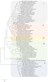
Phylogenetic analysis of the IrlacDyP and other fungal sequences from GenBank (sequences ALD10059.1, AVJ41190, AZJ17937, AZJ17936, AZJ17935, 62271, JQ650250, AAB58908, and ANA52681.1) and Peroxibase database (rest of sequences). Enzymes belonging to subfamilies B and D, and 10222 BaDyPrx01 from subfamily C, were included.
Figure 4
Figure 4
IrlacDyP purification. (A) Elution profile of the enzyme by anion exchange chromatography. (B) SDS–PAGE analysis of samples along the purification procedure. Lane 1, inclusion bodies (10 μg); lane 2, refolding mixture (5 μg); lane 3, purified IrlacDyP (1 μg); lane 4, molecular weight markers.
Figure 5
Figure 5
Spectroscopic characterization of I. lacteus DyP. (A) Electronic absorption spectrum (280–700 nm) of the purified IrlacDyP. (B) Electronic absorption spectra of 2.48 nM IrlacDyP before (black) and after addition of 5 H2O2 equivalents (~12.4 nM) at pH 3 (dark green line). (C) Electronic absorption spectra of 2.48 nM IrlacDyP before (black) and after addition of 5 H2O2 equivalents (~12.4 nM) at pH 5.5 (dark blue line). For (B,C), total spectrum was measured for 30 min at 5 s intervals. (D) Circular dichroism spectrum of the protein.
Figure 6
Figure 6
(A) Optimum pH for purified IrlacDyP. pH stability of IrlacDyP measured as residual activity with ABTS after incubation at different pH values at (B) 4 °C and (C) 25 °C. (D) T50 estimation for purified IrlacDyP.
Figure 7
Figure 7
Hydrogen peroxide stability of IrlacDyP. Residual activity was determined with ABTS.
Figure 8
Figure 8
(A) Superimposition of the I. lacteus DyP (pink) and A. auricula-judae (PDB 4UZI) (yellow) structural models. (B) Heme cofactor environment residues, with Asp172 and Arg335 as putative residues involved in enzyme activation by hydrogen peroxide, His312 occupying the fifth coordination position of the heme iron; solvent-exposed Trp380 and Tyr340 (homologous to AauDyP catalytic Trp377 and protein radical forming Tyr337) are also shown. The white arrow indicates heme channel entrance.
Figure 9
Figure 9
Analysis of phenolic content of softwood (A) and hardwood LS (B) in the presence of 0.25 μM and 0.50 μM of IrlacDyP and 40 and 80 mM H2O2, respectively, after 24 h of treatment. Controls without enzyme and H2O2 are included as grey bars.

References

    1. Beckham G.T., Johnson C.W., Karp E.M., Salvachúa D., Vardon D.R. Opportunities and challenges in biological lignin valorization. Curr. Opin. Biotechnol. 2016;42:40–53. doi: 10.1016/j.copbio.2016.02.030. - DOI - PubMed
    1. Boerjan W., Ralph J., Baucher M. Lignin Biosynthesis. Annu. Rev. Plant Biol. 2003;54:519–546. doi: 10.1146/annurev.arplant.54.031902.134938. - DOI - PubMed
    1. Ruiz-Dueñas F.J., Martínez Á.T. Microbial degradation of lignin: How a bulky recalcitrant polymer is efficiently recycled in nature and how we can take advantage of this. Microb. Biotechnol. 2009;2:164–177. doi: 10.1111/j.1751-7915.2008.00078.x. - DOI - PMC - PubMed
    1. Martínez Á.T., Speranza M., Ruiz-Dueñas F.J., Ferreira P., Camarero S., Guillén F., Martínez M.J., Gutiérrez A., Del Río J.C. Biodegradation of lignocellulosics: Microbial, chemical, and enzymatic aspects of the fungal attack of lignin. Int. Microbiol. 2005;8:195–204. - PubMed
    1. Martínez A.T., Ruiz-Dueñas F.J., Camarero S., Serrano A., Linde D., Lund H., Vind J., Tovborg M., Herold-Majumdar O.M., Hofrichter M., et al. Oxidoreductases on their way to industrial biotransformations. Biotechnol. Adv. 2017;35:815–831. doi: 10.1016/j.biotechadv.2017.06.003. - DOI - PubMed